

Secretagogin is expressed in sensory CGRP neurons and in spinal cord of mouse and complements other calcium-binding proteins, with a note on rat and human
- PMID: 23102406
- PMCID: PMC3560279
- DOI: 10.1186/1744-8069-8-80
Secretagogin is expressed in sensory CGRP neurons and in spinal cord of mouse and complements other calcium-binding proteins, with a note on rat and human
Abstract
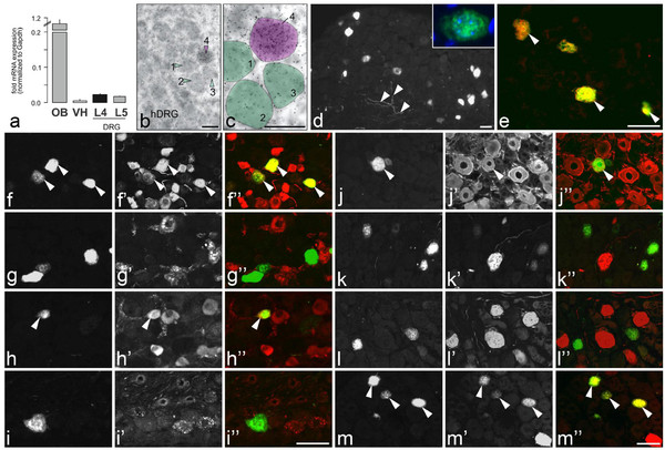
Background: Secretagogin (Scgn), a member of the EF-hand calcium-binding protein (CaBP) superfamily, has recently been found in subsets of developing and adult neurons. Here, we have analyzed the expression of Scgn in dorsal root ganglia (DRGs) and trigeminal ganglia (TGs), and in spinal cord of mouse at the mRNA and protein levels, and in comparison to the well-known CaBPs, calbindin D-28k, parvalbumin and calretinin. Rat DRGs, TGs and spinal cord, as well as human DRGs and spinal cord were used to reveal phylogenetic variations.
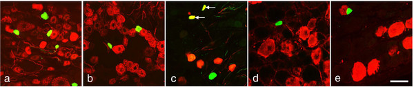
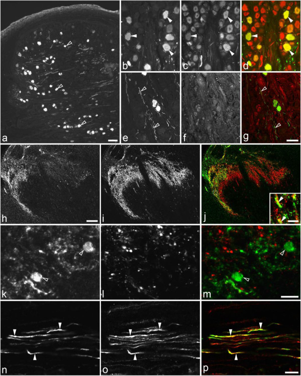
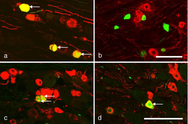
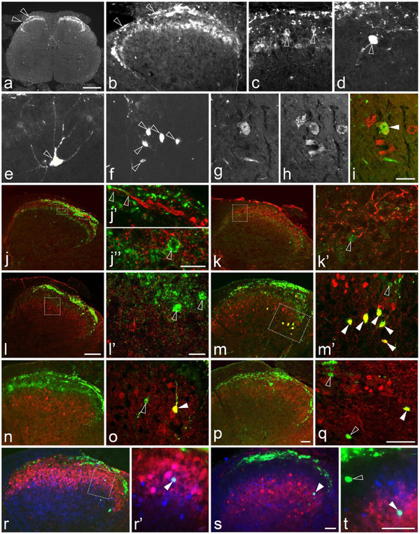
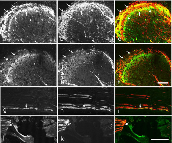
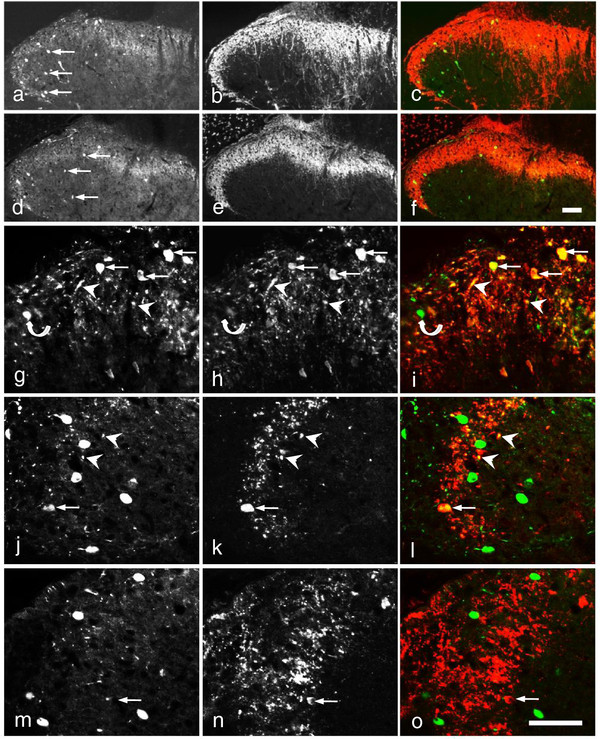
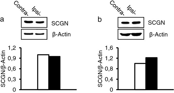
Results: We found Scgn mRNA expressed in mouse and human DRGs and in mouse ventral spinal cord. Our immunohistochemical data showed a complementary distribution of Scgn and the three CaBPs in mouse DRG neurons and spinal cord. Scgn was expressed in ~7% of all mouse DRG neuron profiles, mainly small ones and almost exclusively co-localized with calcitonin gene-related peptide (CGRP). This co-localization was also seen in human, but not in rat DRGs. Scgn could be detected in the mouse sciatic nerve and accumulated proximal to its constriction. In mouse spinal cord, Scgn-positive neuronal cell bodies and fibers were found in gray matter, especially in the dorsal horn, with particularly high concentrations of fibers in the superficial laminae, as well as in cell bodies in inner lamina II and in some other laminae. A dense Scgn-positive fiber network and some small cell bodies were also found in the superficial dorsal horn of humans. In the ventral horn, a small number of neurons were Scgn-positive in mouse but not rat, confirming mRNA distribution. Both in mouse and rat, a subset of TG neurons contained Scgn. Dorsal rhizotomy strongly reduced Scgn fiber staining in the dorsal horn. Peripheral axotomy did not clearly affect Scgn expression in DRGs, dorsal horn or ventral horn neurons in mouse.
Conclusions: Scgn is a CaBP expressed in a subpopulation of nociceptive DRG neurons and their processes in the dorsal horn of mouse, human and rat, the former two co-expressing CGRP, as well as in dorsal horn neurons in all three species. Functional implications of these findings include the cellular refinement of sensory information, in particular during the processing of pain.
Figures
Similar articles
-
G protein-gated inwardly rectifying potassium channel subunits 1 and 2 are down-regulated in rat dorsal root ganglion neurons and spinal cord after peripheral axotomy.Mol Pain. 2015 Jul 22;11:44. doi: 10.1186/s12990-015-0044-z. Mol Pain. 2015. PMID: 26199148 Free PMC article.
-
Phenotyping of sensory and sympathetic ganglion neurons of a galanin-overexpressing mouse--possible implications for pain processing.J Chem Neuroanat. 2006 Jun;31(4):243-62. doi: 10.1016/j.jchemneu.2006.02.001. Epub 2006 Mar 20. J Chem Neuroanat. 2006. PMID: 16546349
-
Cellular localization of three vesicular glutamate transporter mRNAs and proteins in rat spinal cord and dorsal root ganglia.Synapse. 2003 Nov;50(2):117-29. doi: 10.1002/syn.10249. Synapse. 2003. PMID: 12923814
-
A comparative study of the calcium-binding proteins calbindin-D28K, calretinin, calmodulin and parvalbumin in the rat spinal cord.Brain Res Brain Res Rev. 1994 May;19(2):163-79. doi: 10.1016/0165-0173(94)90010-8. Brain Res Brain Res Rev. 1994. PMID: 8061685 Review.
-
Calcium-binding protein parvalbumin in the spinal cord and dorsal root ganglia.Neurochem Int. 2023 Dec;171:105634. doi: 10.1016/j.neuint.2023.105634. Epub 2023 Nov 13. Neurochem Int. 2023. PMID: 37967669
Cited by
-
Ca2+-binding protein NECAB2 facilitates inflammatory pain hypersensitivity.J Clin Invest. 2018 Aug 31;128(9):3757-3768. doi: 10.1172/JCI120913. Epub 2018 Jul 30. J Clin Invest. 2018. PMID: 29893745 Free PMC article.
-
Functional Differentiation of Cholecystokinin-Containing Interneurons Destined for the Cerebral Cortex.Cereb Cortex. 2017 Apr 1;27(4):2453-2468. doi: 10.1093/cercor/bhw094. Cereb Cortex. 2017. PMID: 27102657 Free PMC article.
-
Neuronal calcium-binding proteins 1/2 localize to dorsal root ganglia and excitatory spinal neurons and are regulated by nerve injury.Proc Natl Acad Sci U S A. 2014 Mar 25;111(12):E1149-58. doi: 10.1073/pnas.1402318111. Epub 2014 Mar 10. Proc Natl Acad Sci U S A. 2014. PMID: 24616509 Free PMC article.
-
Developmental and adult characterization of secretagogin expressing amacrine cells in zebrafish retina.PLoS One. 2017 Sep 26;12(9):e0185107. doi: 10.1371/journal.pone.0185107. eCollection 2017. PLoS One. 2017. PMID: 28949993 Free PMC article.
-
The translatability of pain across species.Philos Trans R Soc Lond B Biol Sci. 2019 Nov 11;374(1785):20190286. doi: 10.1098/rstb.2019.0286. Epub 2019 Sep 23. Philos Trans R Soc Lond B Biol Sci. 2019. PMID: 31544615 Free PMC article. Review.
References
-
- Celio RC, Pauls T, Schwaller B. Guidebook to the calcium-binding proteins. New York: Oxford University Press; 1996.
-
- Celio MR. Calbindin D-28k and parvalbumin in the rat nervous system. Neuroscience. 1990;35:375–475. - PubMed
-
- Freund TF, Buzsaki G. Interneurons of the hippocampus. Hippocampus. 1996;6:347–470. - PubMed
-
- Celio MR, Heizmann CW. Calcium-binding protein parvalbumin as a neuronal marker. Nature. 1981;293:300–302. - PubMed
-
- Andressen C, Blumcke I, Celio MR. Calcium-binding proteins: selective markers of nerve cells. Cell Tissue Res. 1993;271:181–208. - PubMed
Publication types
MeSH terms
Substances
Grants and funding
LinkOut - more resources
Full Text Sources
Molecular Biology Databases
Research Materials
Miscellaneous
