. 2012 Oct 26:12:195.
doi: 10.1186/1471-2229-12-195.

Plant plasma membrane-bound staphylococcal-like DNases as a novel class of eukaryotic nucleases

Affiliations

Plant plasma membrane-bound staphylococcal-like DNases as a novel class of eukaryotic nucleases

Krzysztof Leśniewicz et al. BMC Plant Biol. .

Abstract

Background: The activity of degradative nucleases responsible for genomic DNA digestion has been observed in all kingdoms of life. It is believed that the main function of DNA degradation occurring during plant programmed cell death is redistribution of nucleic acid derived products such as nitrogen, phosphorus and nucleotide bases. Plant degradative nucleases that have been studied so far belong mainly to the S1-type family and were identified in cellular compartments containing nucleic acids or in the organelles where they are stored before final application. However, the explanation of how degraded DNA components are exported from the dying cells for further reutilization remains open.

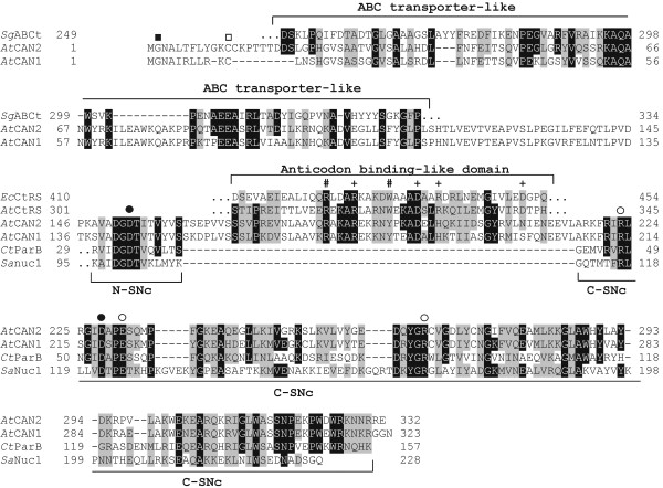
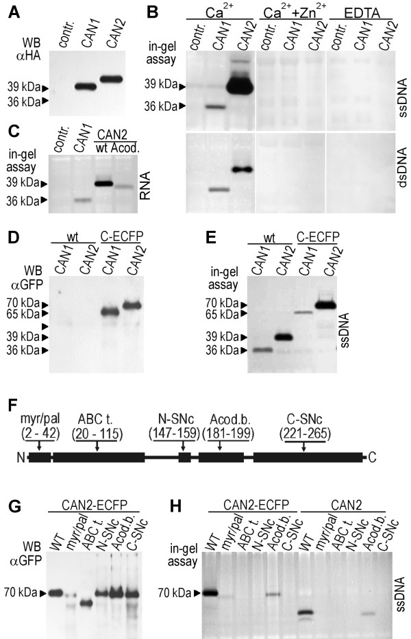
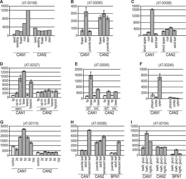
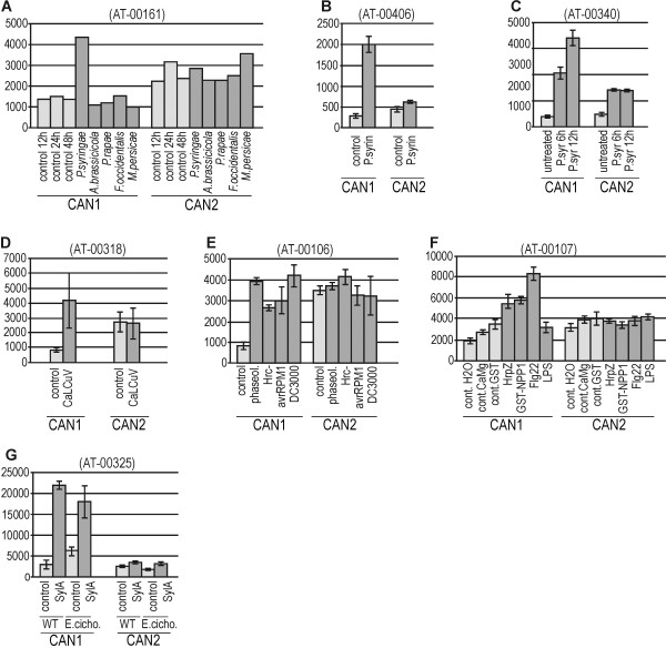
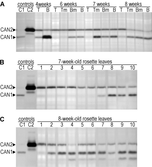
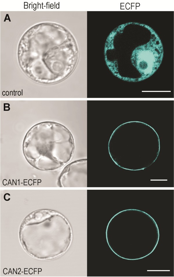
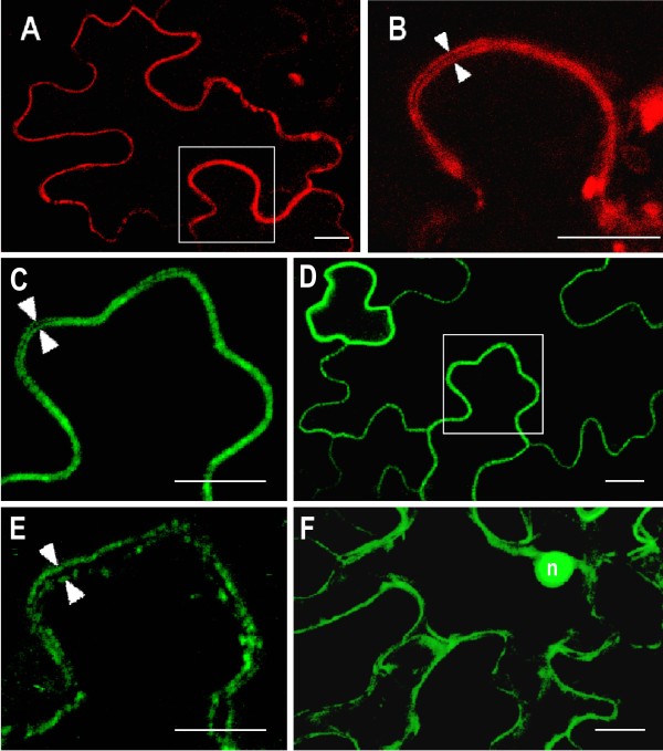
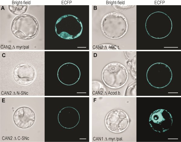
Results: Bioinformatic and experimental data presented in this paper indicate that two Arabidopsis staphylococcal-like nucleases, named CAN1 and CAN2, are anchored to the cell membrane via N-terminal myristoylation and palmitoylation modifications. Both proteins possess a unique hybrid structure in their catalytic domain consisting of staphylococcal nuclease-like and tRNA synthetase anticodon binding-like motifs. They are neutral, Ca2+-dependent nucleaces showing a different specificity toward the ssDNA, dsDNA and RNA substrates. A study of microarray experiments and endogenous nuclease activity revealed that expression of CAN1 gene correlates with different forms of programmed cell death, while the CAN2 gene is constitutively expressed.

Conclusions: In this paper we present evidence showing that two plant staphylococcal-like nucleases belong to a new, as yet unidentified class of eukaryotic nucleases, characterized by unique plasma membrane localization. The identification of this class of nucleases indicates that plant cells possess additional, so far uncharacterized, mechanisms responsible for DNA and RNA degradation. The potential functions of these nucleases in relation to their unique intracellular location are discussed.


Figures

Figure 1
Figure 1
Domain organization and sequence alignments of Arabidopsis CAN1 and CAN2 proteins. The amino acid sequence alignment of two Arabidopsis CAN nucleases with conserved motifs of five bacterial proteins. The alignment contains the full-length sequences of CAN1 and CAN2 proteins and parts of the bacterial proteins showing the highest similarity to the corresponding regions of the CAN1 and CAN2 sequences (brackets above and below the sequences). AtCAN1 – CAN1 nuclease of A. thaliana [TAIR:At3g56170]. AtCAN2 – CAN2 nuclease of A. thaliana [TAIR:At2g40410]. SgABCt - putative periplasmic component of an ABC-type transporter of Syntrophobotulus glycolicus [NCBI:YP_004267097]. EcCtRS - cysteinyl-tRNA synthetase of Escherichia coli [GenBank:EGW73615]. AtCtRS – putative cysteinyl-tRNA synthetase of Anaerolinea thermophila [NCBI:YP_004174706]. CtParB - parB-like nuclease of Cronobacter turicensis [NCBI:YP_003212717]. SaNuc1 - thermonuclease of Staphylococcus aureus [GenBank:EGS91983]. Identical and highly conserved amino acids are boxed in black and gray, respectively. Black square (■) - the N-terminal glycine in the putative N-myristoylation motif. Open square (□) - cysteine residues in the putative palmitoylation motif. Plus sign (+) – residues, which in cysteinyl-tRNA synthetases interact with anticodon-loop bases of bacterial tRNACys. Hash sign (#) residues affecting anticodon binding stability. Black dots () - residues involved in binding of Ca2+ ions. Open dots (○) - residues within the active site of the SNase domain. N-SNc and C-SNc – N-terminal and C-terminal parts of a putative SNase catalytic domain, respectively.
Figure 2
Figure 2
Detection of transiently expressed CAN1 and CAN2 nuclease activity. The protein extract from protoplasts transformed with empty vectors (pSAT6A-ECFP-N1) was used as a negative control (contr.). (A) Western blot analysis of HA-tagged CAN1 and CAN2 nucleases with anti-HA antibody. Molecular mass deduced from gel electrophoresis as indicated by the arrowheads. (B) Detection of CAN1 and CAN2 DNase activities using the in-gel nuclease activity assay. Type of DNA substrate (ssDNA, dsDNA), ions and EDTA added to the reaction buffers, as indicated. (C) RNase activities of CAN1 wild type and CAN2 wild type (wt) and its mutant lacking anti-codon binding-like domain (Acod) are shown. (D) Western blot analysis of wild type (wt) and ECFP-tagged (C-ECFP) CAN1 and CAN2 proteins with anti-GFP antibody. (E) Detection of wild type (wt) and C-terminal ECFP-tagged (C-ECFP) CAN1 and CAN2 DNase activity using in-gel nuclease activity assay. (F) The deletion mutants of CAN2 lacking the conserved regions of domains described in the text. (Myr/pal.) - myristoylation and palmitoylation motif. (ABC t.) - ABC transporter-like sequence. (N-SNc) - N-terminal part of SNc domain. (C-SNc) - C-terminal part of SNc domain. (Acod.b.) - anti-codon binding-like domain. Numbers in the brackets below the domain name abbreviations indicate the ranges of amino acids removed from each mutant. (G) Western blot analysis of CAN2 mutants with an anti-GFP antibody. The abbreviations of the mutated domains as above (Figure  2F). (H) Detection of the nuclease activity of CAN2 mutants. The abbreviations of mutated domains as above (Figure  2F).
Figure 3
Figure 3
Microarray-based expression analysis of CAN1 and CAN2 genes at different developmental stages of Arabidopsis thaliana. Microarray expression data were retrieved from the Genevestigator database and processed as described in the Methods section. Numbers in brackets refer to the experiment ID from Genevestigator. Expression levels of genes encoding CAN1 [TAIR:At3g56170] and CAN2 [TAIR:At2g40410] proteins are displayed as signal values (y-axis) calculated for both genes in a given experiment. The x-axis indicates treatment conditions as described in the text and below. Light gray bars – controls, dark gray bars – expression in response to treatments within a given experiment. (A-C) Three examples of experiments demonstrating predominant expression of the CAN1 gene in Arabidopsis stem. (D-E) Two experiments showing the dependence of CAN1 expression on the stem growth phase. The abbreviations shown on the x-axis indicate the parts of stem: (tip) – stem tip, (up.mi.) - upper middle part of stem, (lo.mi.)- lower middle part of stem, (base) - stem base. (cov) - A. thaliana cov mutant affecting the vascular tissue development. (F-G) Correlation of CAN1 expression with xylem development in the root vascular bundle (F) and in cells induced to differentiate into tracheary elements (G). The abbreviations (2d-10d) indicate the number of days after culture induction. (H-I) correlation of CAN1 expression level with age-dependent (H) and sugar treatment induced senescence (I). The abbreviations (low/high N) and (gluc +/−) indicate the nitrogen and glucose concentration, respectively, in the culture medium. Expression profiles of senescence-associated BFN1 nuclease are also shown.
Figure 4
Figure 4
Microarray-based expression analysis of CAN1 and CAN2 genes in response to various stimuli associated with pathogenesis. Microarray expression data were retrieved from the Genevestigator database and processed as described in the Methods section. Numbers in brackets refer to the experiment ID from Genevestigator. Expression levels of genes encoding CAN1 [TAIR:At3g56170] and CAN2 [TAIR:At2g40410] proteins are displayed as signal values (y-axis) calculated for both genes in a given experiment. The x-axis indicates treatment conditions as described in the text and below. Light gray bars – controls, dark gray bars – expression in response to treatments within a given experiment. Detailed references to individual experiments are given in Additional file 2. (A) Microarray study showing specific activation of CAN1 expression in response to bacterial infection. Plant responses to the following microbial and insect pathogens are presented: leaf bacterium (Pseudomonas syringae pv. tomato), pathogenic leaf fungus (Alternaria brassicicola), tissue-chewing caterpillars (Pieris rapae), cell-content-feeding thrips (Frankliniella occidentalis) and phloem-feeding aphids (Myzus persicae). (B-D) Three experiments showing the effect of P. syringae (B-C) and DNA virus (cabbage leaf curl virus-CaLCuV) (D) on CAN1 gene expression. (E) Influence of different P. syringae strains on CAN1 expression. (F) Influence of different pathogen-derived elicitors on CAN1 gene expression. (HrpZ) - Harpin elicitor, (NPP1) - necrosis-inducing Phytophthora protein 1, (Flg22) – flagellin, (LPS) – lipopolysaccharide. (G) Expression of CAN1 gene in plants treated with Syringolin A (SylA) alone and with a combination of Syringolin A and Erysiphe cichoracearum infection.
Figure 5
Figure 5
Endogenous CAN1 and CAN2 activity. (A) Changes in endogenous CAN1 and CAN2 activity during stem development. DNase activity of protein extracts from the 4–8 week old stems were analyzed. The abbreviations for the gel tracks refer to the part of stem: (T) – stem tip, (Tm) - upper middle part of stem, (Bm) - lower middle part of stem, (B) - stem base. CAN1 (C1) and CAN2 (C1) nucleases transiently expressed in protoplasts were used as positive controls. (B-C) The endogenous CAN1 and CAN2 nuclease activity of individual leaves of 7 week old (B) and 8 week old (C) rosettes. Consecutive leaf extracts are arranged from the youngest (1) to oldest (10) leaf of rosettes. The CAN1 (C1) and CAN2 (C1) controls as above (Figure  5A).
Figure 6
Figure 6
Plasma membrane localization of CAN1 and CAN2 nucleases in protoplasts. ECFP alone and ECFP-fusion proteins were transiently expressed in Arabidopsis root cell protoplasts. Localization was analyzed by confocal laser scanning microscopy (right half of each panel). The corresponding bright-field images are shown on the left halves of panels. (A) Protoplast transformed with empty vector (pSAT6A-ECFP) as a control shows characteristic cytoplasmatic and nuclear localization (B) Expression of the CAN1-ECFP fusion construct. (C) Expression of the CAN2-ECFP fusion construct. Clear plasma membrane localization of both nucleases is seen. The length of the white scale bar at the right bottom of each panel corresponds to 10 μm.
Figure 7
Figure 7
Plasma membrane localization of CAN1-RFP and CAN2-GFP nucleases transiently expressed in Arabidopsis leaf epidermal cells. Confocal images were taken two days after A. tumefaciens infiltration. (A) An epidermal cell expressing CAN1-RFP fusion protein. (B) Higher magnification image of the box in (A). White arrowheads indicate plasma membranes of adjacent cells. (C-E) Epidermal cells expressing the CAN2-GFP fusion protein. (C and E) Higher magnification images of the box in (D) show plasma membranes of adjacent cells before (C) and after (D) plasmolysis. (F) Epidermal cells transformed with empty vector pSITE-2NB-GFP as a control. Strong signal around the nucleus (n) and numerous cytoplasmic strands show characteristic cytoplasmatic localization of GFP protein. The length of the white scale bar at the right bottom of each panel corresponds to 10 μm.
Figure 8
Figure 8
Subcellular localization of CAN nucleases deletion mutants. ECFP-fusion proteins were transiently expressed in Arabidopsis root cell protoplasts. The bright-field images are shown in the left part of each panel and corresponding confocal laser scanning images are on the right. The images show the cellular location of the ECFP fused proteins lacking the following consensus sequences: (A) CAN2 mutant lacking N-myristoylation and palmitoylation consensus sequences. (B) CAN2 mutant lacking the ABC transporter-like sequence. (C) CAN2 mutant lacking the N-SNc domain. (D) CAN2 mutant lacking the anticodon binding-like sequence (E) CAN2 mutant lacking the C-SNc domain. (F) CAN1 mutant lacking N-myristoylation and palmitoylation consensus sequences. The abbreviations of mutated domains as in Figure  2F The length of the white scale bar at the right bottom of each panel corresponds to 10 μm.

Similar articles

Cited by

References

    1. Nagata S, Nagase H, Kawane K, Mukae N, Fukuyama H. Degradation of chromosomal DNA during apoptosis. Cell Death Differ. 2003;10:108–16. doi: 10.1038/sj.cdd.4401161. - DOI - PubMed
    1. Thomas H, Ougham HJ, Wagstaff C, Stead AD. Defining senescence and death. J Exp Bot. 2003;54:1127–32. doi: 10.1093/jxb/erg133. - DOI - PubMed
    1. Guo Y, Cai Z, Gan S. Transcriptome of Arabidopsis leaf senescence. Plant Cell Environ. 2004;27:521–549. doi: 10.1111/j.1365-3040.2003.01158.x. - DOI
    1. Aoyagi S, Sugiyama M, Fukuda H. BEN1 and ZEN1 cDNAs encoding S1-type DNases that are associated with programmed cell death in plants. FEBS Lett. 1998;429:134–8. doi: 10.1016/S0014-5793(98)00563-8. - DOI - PubMed
    1. Pérez-Amador MA, Abler ML, De Rocher EJ, Thompson DM, van Hoof A, LeBrasseur ND, Lers A, Green PJ. Identification of BFN1, a bifunctional nuclease induced during leaf and stem senescence in Arabidopsis. Plant Physiol. 2000;122:169–80. doi: 10.1104/pp.122.1.169. - DOI - PMC - PubMed

Publication types

MeSH terms