. 2012 Nov 13;23(5):1020-31.
doi: 10.1016/j.devcel.2012.09.016. Epub 2012 Oct 25.

Gadd45γ and Map3k4 interactions regulate mouse testis determination via p38 MAPK-mediated control of Sry expression

Affiliations

Gadd45γ and Map3k4 interactions regulate mouse testis determination via p38 MAPK-mediated control of Sry expression

Nick Warr et al. Dev Cell. .

Abstract

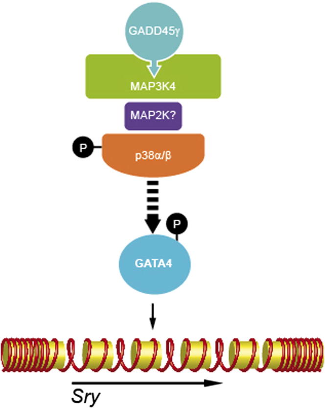
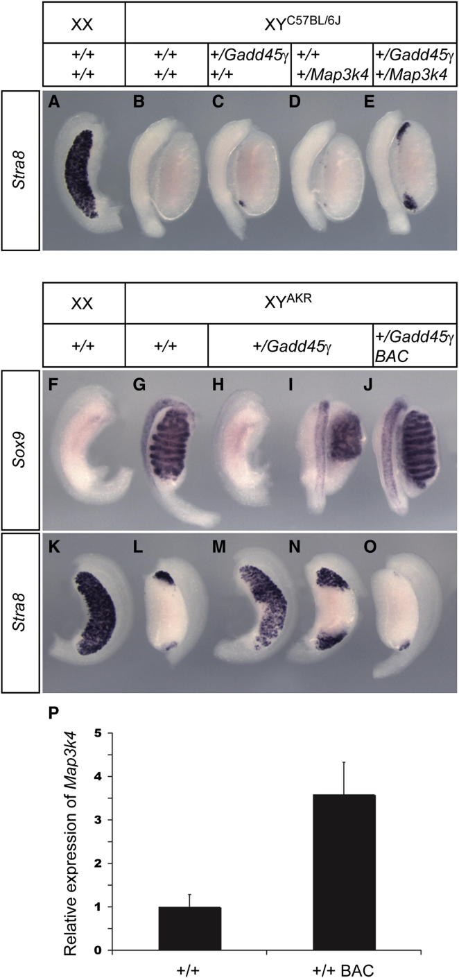
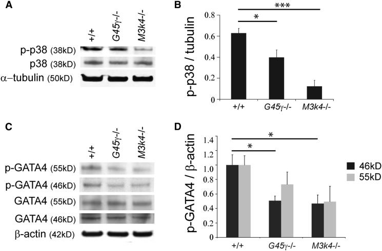
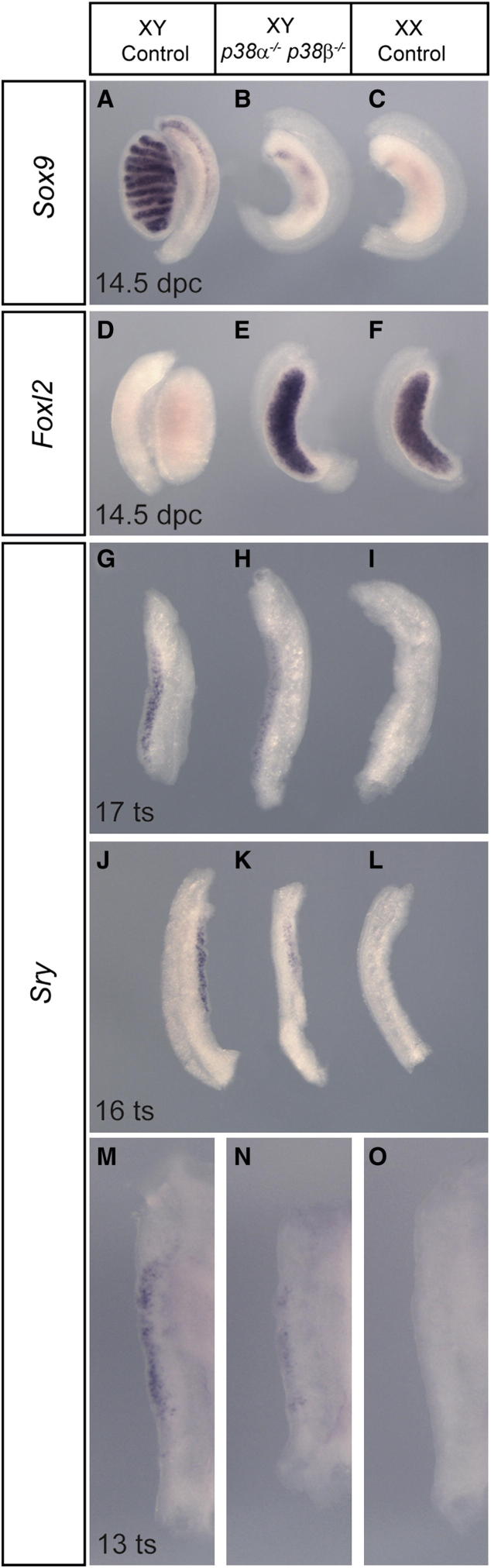
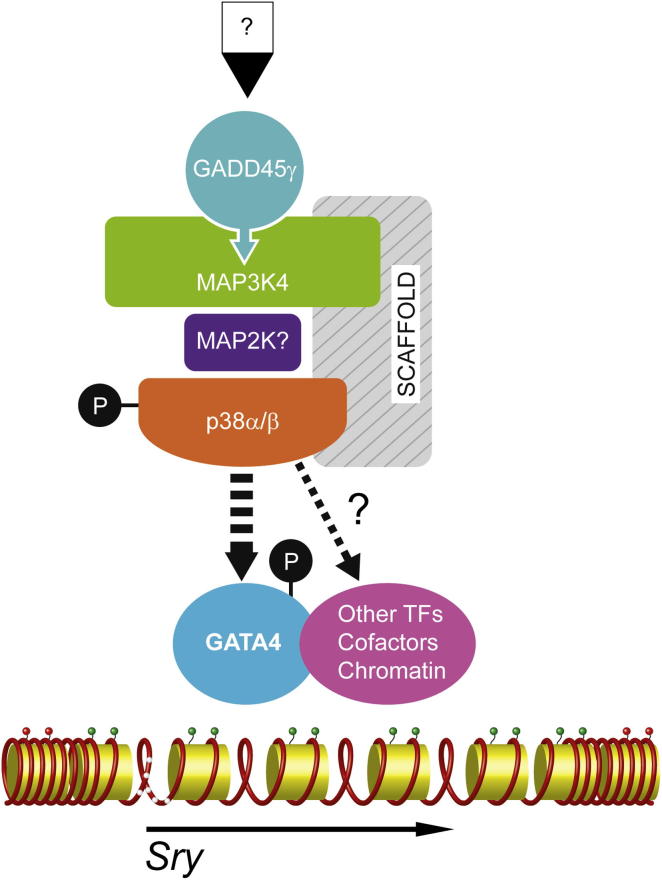
Loss of the kinase MAP3K4 causes mouse embryonic gonadal sex reversal due to reduced expression of the testis-determining gene, Sry. However, because of widespread expression of MAP3K4, the cellular basis of this misregulation was unclear. Here, we show that mice lacking Gadd45γ also exhibit XY gonadal sex reversal caused by disruption to Sry expression. Gadd45γ is expressed in a dynamic fashion in somatic cells of the developing gonads from 10.5 days postcoitum (dpc) to 12.5 dpc. Gadd45γ and Map3k4 genetically interact during sex determination, and transgenic overexpression of Map3k4 rescues gonadal defects in Gadd45γ-deficient embryos. Sex reversal in both mutants is associated with reduced phosphorylation of p38 MAPK and GATA4. In addition, embryos lacking both p38α and p38β also exhibit XY gonadal sex reversal. Taken together, our data suggest a requirement for GADD45γ in promoting MAP3K4-mediated activation of p38 MAPK signaling in embryonic gonadal somatic cells for testis determination in the mouse.


Figures

None
Graphical abstract
Figure 1
Figure 1
Gadd45γ Is Expressed in Somatic Cells of the Developing XX and XY Gonad with a Spatiotemporal Profile Reminiscent of Sry (A–C) WMISH in the developing XX (A) and XY (B) wild-type gonads at 11.5 dpc (19 ts). Expression is restricted to the body of the gonad and not detected in the coelomic epithelium (C, arrowhead). Dotted line in (B) indicates the position of the section shown in (C). Scale bar in (C) is 100 μm. (D and E) Two-color WMISH with Oct4 (red) and Gadd45γ (blue). (F–I) Oct4 is not detected in W/W homozygous gonads at 11.5 dpc (F and G), in contrast to Gadd45γ expression (H and I). (J–L) Gadd45γ is first detected at 9 ts, in the central portion of the gonad (J and K); white arrowheads mark anterior and posterior limits of the gonad in (J–L). By 10 ts expression is detected throughout the gonad (L). (M–T) Gadd45γ is expressed at high levels until around 25 ts (M–R), and declines thereafter, being undetectable at 13.5 dpc (S and T). (U–BB) Sry expression profile in the same genetic background.
Figure 2
Figure 2
XY Mice Lacking Gadd45γ Are Phenotypic Females (A–C) External genitalia of wild-type XX (A), Gadd45γ/ XY (B), and wild-type XY (C) adult mice. (D–F) Mutant XY ovaries (E) appear smaller than their wild-type XX counterparts (D), and some variation in morphology is apparent. Anogenital distance (square brackets) is quantified (F). (G–L) Histological examination reveals that some mutant XY ovaries (H and K), have an almost normal appearance when compared to XX controls (G and J), while others are very hypoplastic with very few or no follicles (I and L). Cysts (asterisks, I and L) were commonly detected in XY mutant ovaries. Scale bar in (G), 1 mm; scale bar in (J), 300 μm. AF, antral follicle; SF, secondary follicle; CL, corpus luteum. Error bars, SEM.
Figure 3
Figure 3
XY Gonadal Sex Reversal in Gadd45γ-Deficient Embryos Is Caused by Disrupted Sry Expression (A–L) Frames containing three gonadal tissue samples have the order, from left to right, XY wild-type, XY Gadd45γ/, and XX wild-type. WMISH reveals reduction in Sry transcripts detected at 11.25 dpc (16 ts) in XY mutants (A). SRY protein levels are similarly diminished in mutants (B, mutant on right). By 12.5 dpc, Sry expression is detectable at higher levels in the mutant XY gonad, in contrast to negligible levels in the XY wild-type (toward posterior of gonad) and XX control at the same stage (C). Sox9 transcripts are undetectable in mutant gonads at 18 ts (D) and 14.5 dpc (F), and SOX9 protein is also not detected at 11.5 dpc (E). AMH is also undetectable in mutant XY gonads at 14.5 dpc (G), and 3βHSD is absent at 13.5 dpc (H). Wnt4 is expressed in mutant XY gonads at 11.5 dpc (I). FOXL2 and Stra8 are expressed at high levels in mutant XY gonads at 14.5 dpc (J and K, respectively), concomitant with downregulation of Oct4 expression (L). (M–O) Loss of Sry and Sox9 expression in XY mutant gonads was confirmed by qRT-PCR (M) at 18 ts, as was inappropriate expression of ovarian marker genes (N). qRT-PCR reveals a delay in Sry expression in Gadd45γ/ and Map3k4/ gonads (O). Error bars, SEM. Dotted white lines mark the boundary between gonad and mesonephros in samples that do not exhibit significant expression of marker protein. See also Figure S1 and Figure S2 for additional phenotypic analyses of Gadd45γ/ and Map3k4/ embryonic gonads.
Figure 4
Figure 4
Genetic Analyses of Functional Interactions between Gadd45γ and Map3k4 during Testis Determination (A and B) Stra8 expression is ovary specific in wild-type gonads at 14.5 dpc. (C–O) Significant numbers of Stra8-positive cells are detectable at poles of XY gonads lacking one copy of both Gadd45γ and Map3k4 (E). Only very few Stra8-positive cells are detected, occasionally, in singly heterozygous gonads (C and D). Sox9 and Stra8 WMISH reveal ovary development (H and M), or ovotestis development (I and N), in embryos lacking a single copy of Gadd45γ on C57BL/6J-YAKR (B6-YAKR), in contrast to wild-type controls (G and L). Wild-type XX littermate controls do not express Sox9 (F) but express high levels of Stra8 (K). The presence of a Map3k4 BAC transgene rescues testis development in Gadd45γ+/− heterozygotes (J and O). (P) Quantitation of gonadal Map3k4 expression by qRT-PCR in wild-type (+/+) and BAC transgenic (+/+ BAC) embryos at 11.5 dpc indicates an approximate 3.5-fold increase of expression in BAC transgenics. Error bars, SEM.
Figure 5
Figure 5
Reduced Phosphorylation of p38 MAPK and GATA4 in Embryonic Gonads Lacking GADD45γ or MAP3K4 (A–C) Immunoblotting of subdissected XY gonad samples at the 15–16 ts stage (A and C). Relative quantitation of phospho-p38 normalized to the α-tubulin loading control (B). (D) Relative quantitation of phospho-GATA4 normalized to the β-actin loading control. Error bars, SEM.
Figure 6
Figure 6
XY Sex Reversal in Embryonic Gonads Lacking Both p38α and p38β MAPKs (A–F) WMISH of XY double-knockout embryos reveals loss of Sox9 expression (A–C) and gain of Foxl2 expression (D–F) in gonads at 14.5 dpc, when compared with control genotypes. (G–O) Sry expression is reduced in doubly mutant XY embryonic gonads at 13 ts (N), 16 ts (K), and 17 ts (H), in comparison to XY controls (M, J, and G, respectively). Control embryos lacked at most one copy of p38a. Such control XY gonads never exhibited sex reversal at 14.5 dpc. However, loss of p38α or p38β alleles in the control gonads used here may possibly reduce the observable differences in Sry levels between control and doubly homozygous gonads.
Figure 7
Figure 7
Summary of Proposed Interactions between GADD45γ, MAP3K4, p38 MAPK, GATA4, and Sry in Testis Determination The signals regulating the expression of Gadd45γ shortly after gonad formation (indicated by a question mark at the top) are unknown. Based on data reported here and described in the supporting literature, we propose that GADD45γ activates MAP3K4 in supporting cell precursors of the developing gonad from around 10.5 dpc. MAP3K4 activates p38α and p38β (indicated by a circled P) through an as yet uncharacterized MAP2K. This MAPK phosphorelay module, perhaps in the context of an unidentified scaffold protein, results in the direct or indirect activation of GATA4 by phosphorylation (dashed arrow) and subsequent expression of Sry in XY supporting cells. In addition to DNA demethylation of its promoter region (white circles on DNA strand), which does not require GADD45γ/MAP3K4, timely Sry expression may require chromatin marks also established by this signal transduction pathway (green circles). Additional targets of this MAPK cascade in the developing gonad are not excluded but remain to be identified, possibly comprising other chromatin-associated transcription factors (TFs), transcriptional cofactors, and chromatin-modifying enzymes that alter chromatin configuration at Sry (and/or other loci).

References

    1. Abell A.N., Jordan N.V., Huang W., Prat A., Midland A.A., Johnson N.L., Granger D.A., Mieczkowski P.A., Perou C.M., Gomez S.M. MAP3K4/CBP-regulated H2B acetylation controls epithelial-mesenchymal transition in trophoblast stem cells. Cell Stem Cell. 2011;8:525–537. - PMC - PubMed
    1. Adams R.H., Porras A., Alonso G., Jones M., Vintersten K., Panelli S., Valladares A., Perez L., Klein R., Nebreda A.R. Essential role of p38alpha MAP kinase in placental but not embryonic cardiovascular development. Mol. Cell. 2000;6:109–116. - PubMed
    1. Albrecht K.H., Eicher E.M. Evidence that Sry is expressed in pre-Sertoli cells and Sertoli and granulosa cells have a common precursor. Dev. Biol. 2001;240:92–107. - PubMed
    1. Barreto G., Schäfer A., Marhold J., Stach D., Swaminathan S.K., Handa V., Döderlein G., Maltry N., Wu W., Lyko F., Niehrs C. Gadd45a promotes epigenetic gene activation by repair-mediated DNA demethylation. Nature. 2007;445:671–675. - PubMed
    1. Beardmore V.A., Hinton H.J., Eftychi C., Apostolaki M., Armaka M., Darragh J., McIlrath J., Carr J.M., Armit L.J., Clacher C. Generation and characterization of p38beta (MAPK11) gene-targeted mice. Mol. Cell. Biol. 2005;25:10454–10464. - PMC - PubMed

Publication types

MeSH terms

Substances