

Dynamic 3-dimensional echocardiographic assessment of mitral annular geometry in patients with functional mitral regurgitation
- PMID: 23103005
- PMCID: PMC5228313
- DOI: 10.1016/j.athoracsur.2012.08.078
Dynamic 3-dimensional echocardiographic assessment of mitral annular geometry in patients with functional mitral regurgitation
Abstract
Background: Mitral valve (MV) annular dynamics have been well described in animal models of functional mitral regurgitation (FMR). Despite this, little if any data exist regarding the dynamic MV annular geometry in humans with FMR. In the current study we hypothesized that 3-dimensional (3D) echocardiography, in conjunction with commercially available software, could be used to quantify the dynamic changes in MV annular geometry associated with FMR.
Methods: Intraoperative 3D transesophageal echocardiographic data obtained from 34 patients with FMR and 15 controls undergoing cardiac operations were dynamically analyzed for differences in mitral annular geometry with TomTec 4D MV Assessment 2.0 software (TomTec Imaging Systems GmbH, Munich, Germany).
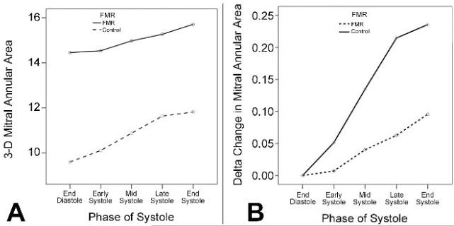
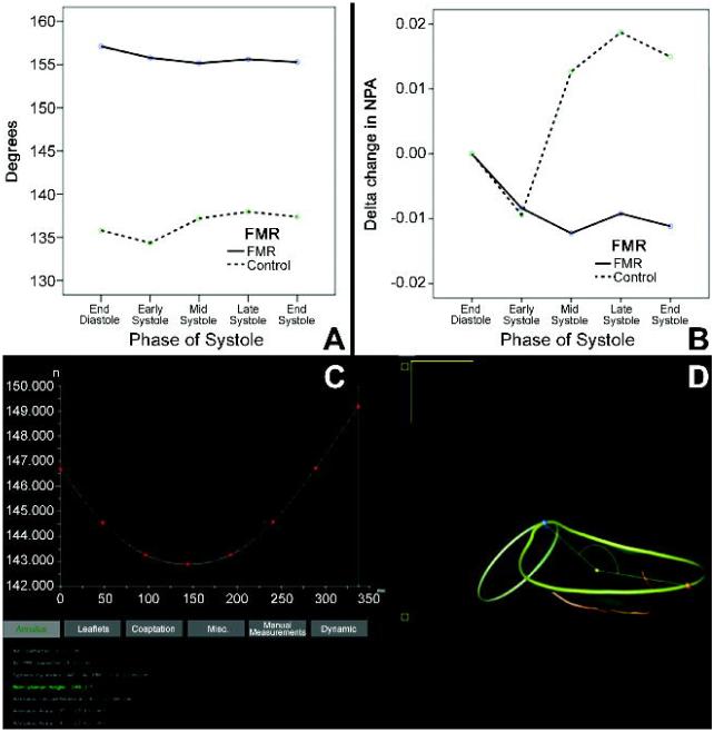
Results: In patients with FMR, the mean mitral annular area (14.6 cm(2) versus 9.6 cm(2)), circumference (14.1 cm versus 11.4 cm), anteroposterior (4.0 cm versus 3.0 cm) and anterolateral-posteromedial (4.3 cm versus 3.6 cm) diameters, tenting volume (6.2 mm(3) versus 3.5 mm(3)) and nonplanarity angle (NPA) (154 degrees ± 15 versus 136 degrees ± 11) were greater at all points during systole compared with controls (p < 0.01). Vertical mitral annular displacement (5.8 mm versus 8.3 mm) was reduced in FMR compared with controls (p < 0.01).
Conclusions: There are significant differences in dynamic mitral annular geometry between patients with FMR and those without. We were able to analyze these changes in a clinically feasible fashion. Ready availability of this information has the potential to aid comprehensive quantification of mitral annular function and possibly assist in both clinical decision making and annuloplasty ring selection.
Copyright © 2013 The Society of Thoracic Surgeons. Published by Elsevier Inc. All rights reserved.
Figures
References
-
- Gorman JH, 3rd, Gupta KB, Streicher JT, et al. Dynamic three-dimensional imaging of the mitral valve and left ventricle by rapid sonomicrometry array localization. The Journal of thoracic and cardiovascular surgery. 1996;112(3):712–26. - PubMed
-
- Daimon M, Saracino G, Fukuda S, et al. Dynamic change of mitral annular geometry and motion in ischemic mitral regurgitation assessed by a computerized 3D echo method. Echocardiography. 2010;27(9):1069–77. - PubMed
-
- Little SH, Ben Zekry S, Lawrie GM, et al. Dynamic annular geometry and function in patients with mitral regurgitation: insight from three-dimensional annular tracking. J Am Soc Echocardiogr. 2010;23(8):872–9. - PubMed
-
- Veronesi F, Corsi C, Sugeng L, et al. Quantification of mitral apparatus dynamics in functional and ischemic mitral regurgitation using real-time 3-dimensional echocardiography. J Am Soc Echocardiogr. 2008;21(4):347–54. - PubMed
Publication types
MeSH terms
Grants and funding
LinkOut - more resources
Full Text Sources
Other Literature Sources
