. 2012 Nov 29;2(5):1259-71.
doi: 10.1016/j.celrep.2012.09.018. Epub 2012 Oct 25.

Cardiovascular protection by ApoE and ApoE-HDL linked to suppression of ECM gene expression and arterial stiffening

Affiliations

Cardiovascular protection by ApoE and ApoE-HDL linked to suppression of ECM gene expression and arterial stiffening

Devashish Kothapalli et al. Cell Rep. .

Abstract

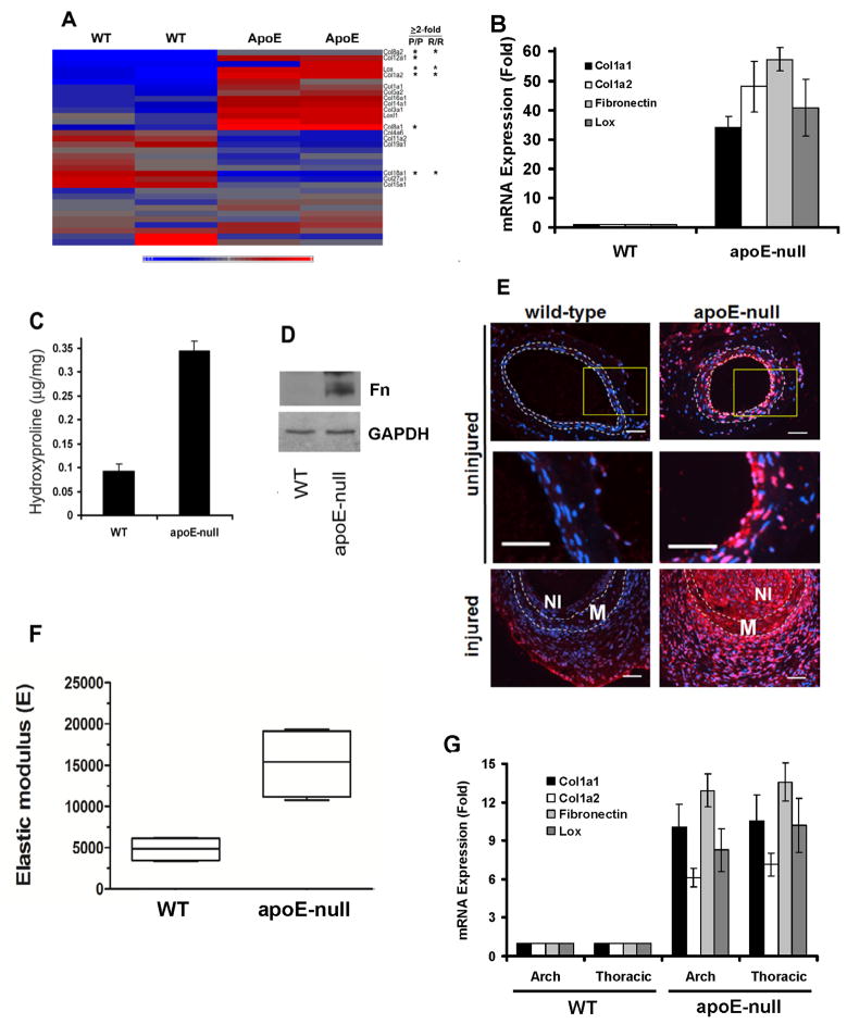
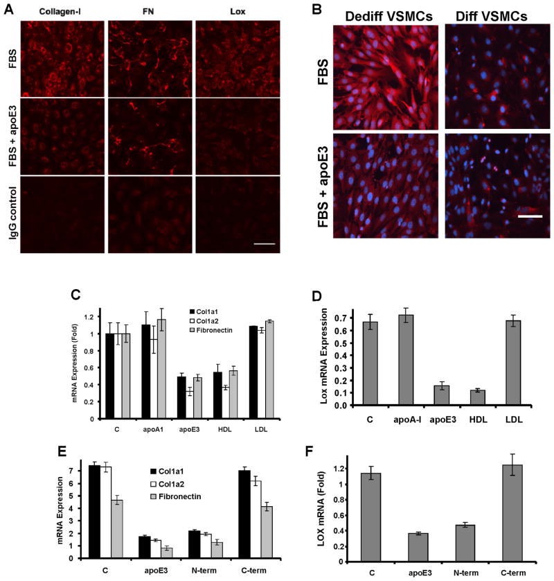
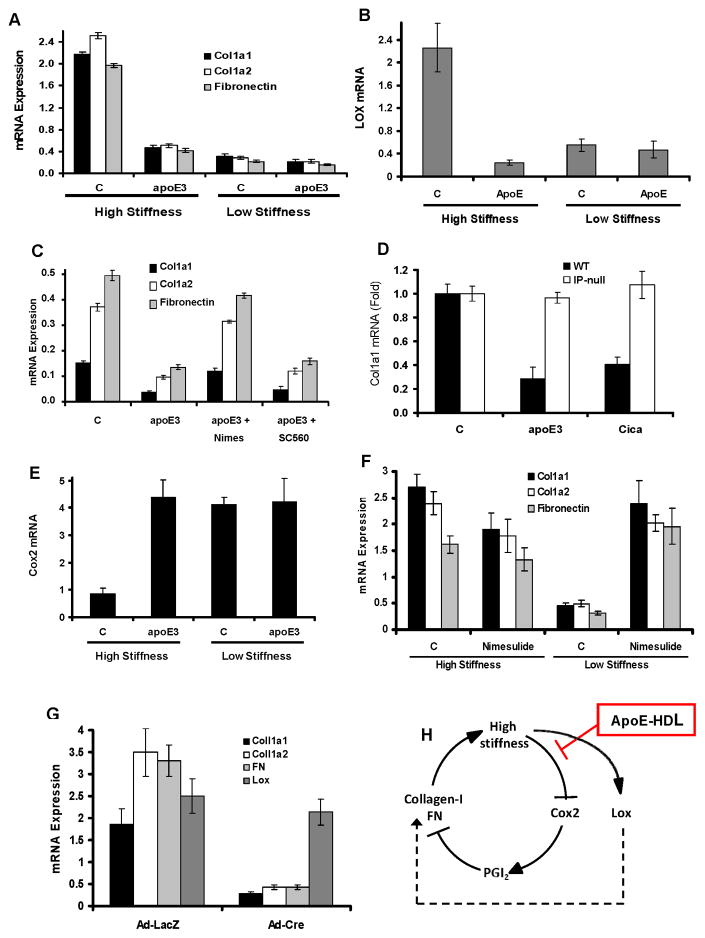
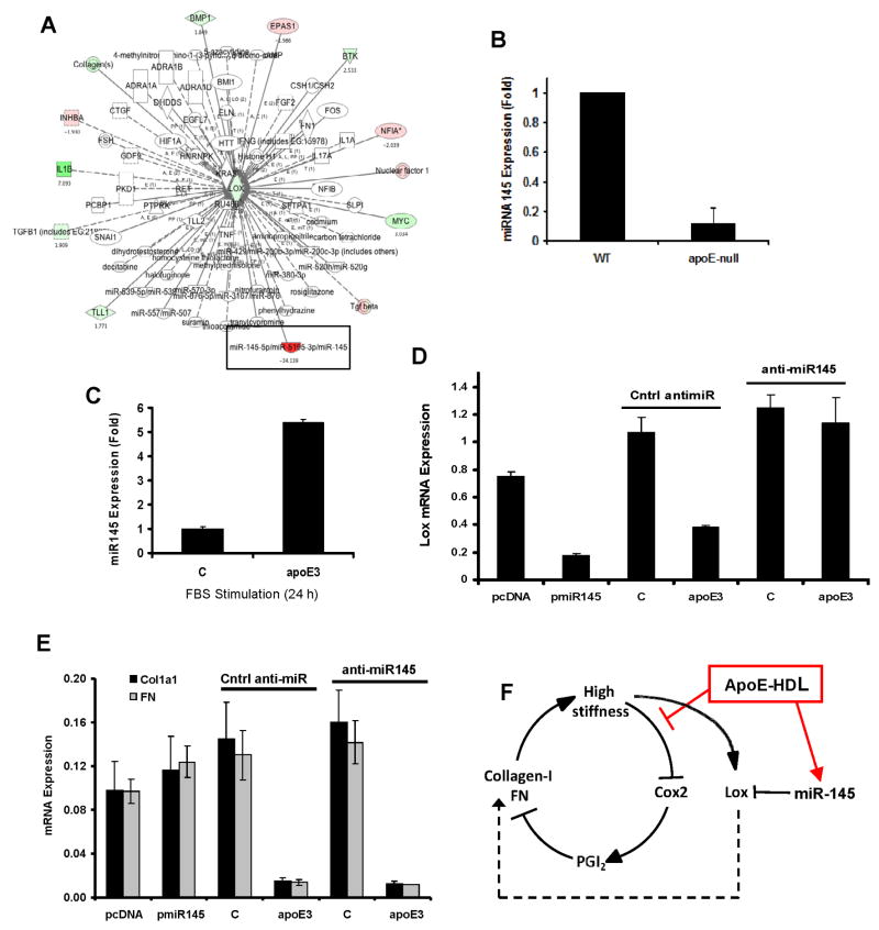
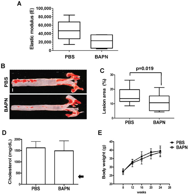
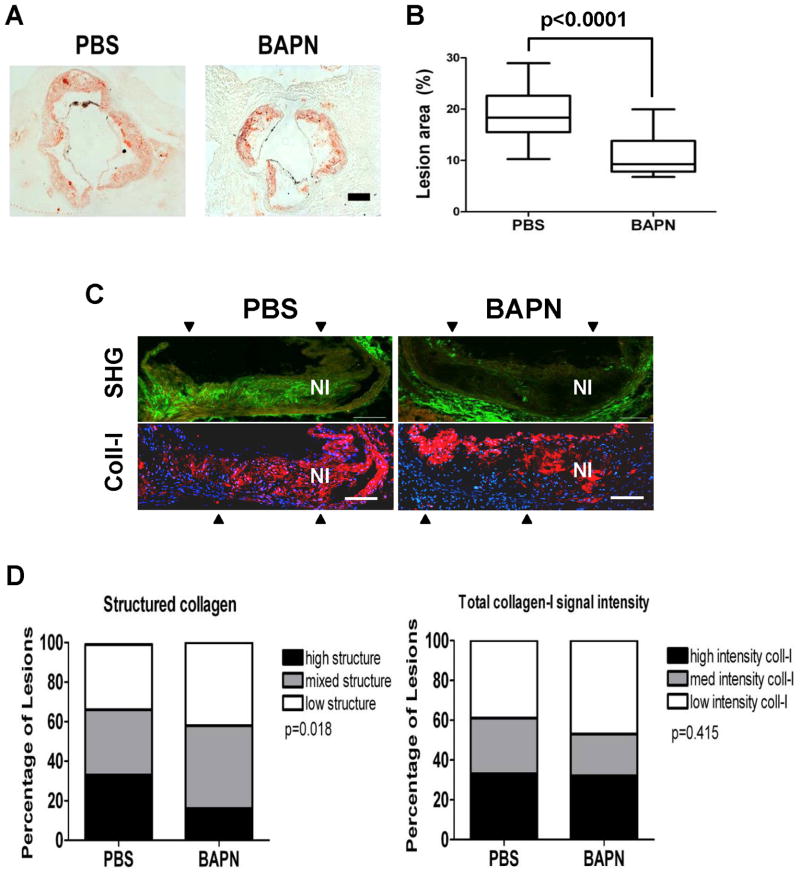
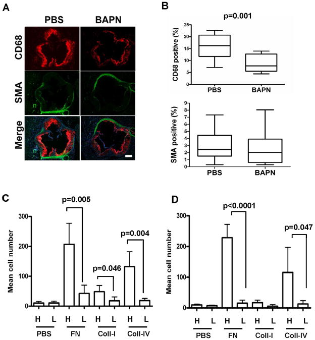
Arterial stiffening is a risk factor for cardiovascular disease, but how arteries stay supple is unknown. Here, we show that apolipoprotein E (apoE) and apoE-containing high-density lipoprotein (apoE-HDL) maintain arterial elasticity by suppressing the expression of extracellular matrix genes. ApoE interrupts a mechanically driven feed-forward loop that increases the expression of collagen-I, fibronectin, and lysyl oxidase in response to substratum stiffening. These effects are independent of the apoE lipid-binding domain and transduced by Cox2 and miR-145. Arterial stiffness is increased in apoE null mice. This stiffening can be reduced by administration of the lysyl oxidase inhibitor BAPN, and BAPN treatment attenuates atherosclerosis despite highly elevated cholesterol. Macrophage abundance in lesions is reduced by BAPN in vivo, and monocyte/macrophage adhesion is reduced by substratum softening in vitro. We conclude that apoE and apoE-containing HDL promote healthy arterial biomechanics and that this confers protection from cardiovascular disease independent of the established apoE-HDL effect on cholesterol.


Figures

Figure 1
Figure 1. Altered ECM gene expression in apoE-null mice
(A) Heat map of collagen and Lox genes in WT and apoE-null aortae. Duplicates represent Agilent dye-swaps. Asterisks indicate fold-change ≥2 in athero-prone (P/P) and resistant (R/R) regions. Scale: −0.9–1.0. (B) Cleaned aortae from 6-mo male WT and apoE-null mice analyzed by RT-qPCR. Results show mean ± SE, n=4. P-0.012 by 2-tailed t-test. (C–D) Cleaned aortae analyzed for hydroxyproline content (mean ± SD, n=3) or western blotted for FN, respectively. (E) Lox immunostaining (red) of uninjured and injured femoral arteries. DAPI-stained nuclei are shown in blue. Dashed lines show the IEL and EEL. M; media. NI; neointima. Middle panels show enlargements of boxed regions. Scale bar = 50 μm. (F) Thoracic aortae of four 6-mo WT and four apoE-null mice were cleaned of adventitia, opened longitudinally, and analyzed by AFM, indenting into the luminal surface at >20 distinct non-lesioned locations. The mean elastic modulus was calculated for each mouse and graphed as a Tukey box and whisker plot; p=0.029 by 2-tailed Mann-Whitney test. (G) Cleaned aortae from 6-mo male WT and apoE-null mice were divided into arch (ascending) and thoracic (descending) regions. RNA was isolated from the aortae and analyzed by RT-qPCR for collagen-I, FN, and Lox gene expression. Results are normalized to 18S rRNA. Results show mean ± SE, n=5. p=0.036 by 2-tailed t-test. See also Figure S1 and Table S1A–C.
Figure 2
Figure 2. ApoE and HDL inhibit ECM expression in dedifferentiated VSMCs
(A) Serum-starved VSMCs isolated from wild-type mice were incubated with 10% FBS in the absence (control; C) or presence of 2 μM apoE3 for 24 h. The cells were fixed and stained for collagen-I, FN or Lox and visualized by immunofluorescence microscopy. Scale bar=50μm. The IgG control used FBS-stimulated cells. (B) VSMCs were isolated from C57BL/6 aortae by explant cultures (dedifferentiated phenotype) or by collagenase digestion (differentiated phenotype). Asynchronous cells of each phenotype were incubated with 10% FBS in the absence (control; C) or presence of 2 μM apoE3 for 24 h and then stained for collagen-I (red). Blue: dapi-stained nuclei. Scale bar=50μm. The figure shows a representative result. (C–D) Serum-starved VSMCs isolated from WT mice were incubated with 10% FBS in the absence (control, C) or presence of 2 μM apoA-I or apoE3 or 50 μg/ml HDL or LDL for 24 h. RNA levels were determined by RT-qPCR. (E–F) The experiment in C–D was repeated except we compared the effects of 2 μM apoE3 to its N- and C-terminal fragments. RT-qPCR results show mean ± SD of duplicate PCR reactions and are representative of at least three independent experiments. See also Figure S2.
Figure 3
Figure 3. Mechano-sensitive gene expression regulated by apoE
(A–B) Serum-starved primary mouse VSMCs were incubated with 10% FBS in the absence (control, C) or presence of 2 μM apoE3 on high stiffness or low stiffness FN-coated hydrogels for 24 h. (C) the experiment in A–B was repeated except that apoE-treated VSMCs on plastic were given 1 μM nimesulide (Nimes; Cox2 inhibitor) or 1 mM SC560 (Cox1 inhibitor). (D) The experiment in C was repeated using WT and IP-null VSMCs treated with 2 μM apoE3 or 200 nM cicaprost (Cica; stable PGI2 mimetic). (E) The experiment in A was analyzed for Cox2 mRNA. (F) Serum-starved primary mouse VSMCs were incubated with 10% FBS in the absence (control, C) or presence of 1 μM nimesulide on high stiffness or low stiffness FN-coated hydrogels for 24 h. (G) Primary VSMCs were isolated from mice expressing Cre-dependent Cox2. The cells were seeded overnight on FN-coated cover slips, serum-starved, infected with adenoviruses encoding LacZ or Cre, and directly stimulated with 10% FBS for 24 h. For all panels, Col1a1, Col1a2, FN, Lox, or Cox2 mRNAs were quantified by RT-qPCR and plotted relative to 18S rRNA. For panels A–G, results show mean ± SD of duplicate PCR reactions and are representative of at least three independent experiments. (H) Model of the stiffness- and apoE-regulatory effects on Cox2, collagen-I, FN, and Lox expression. See also Figure S3.
Figure 4
Figure 4. miR145-dependent Lox gene expression by apoE
(A) Upstream regulators of Lox showing differential gene expression after vascular injury as determined by Ingenuity Pathway Analysis. Green and red represent induction and repression, respectively. (B) Aortae from 6-mo old WT or apoE-null mice were harvested and analyzed by RT-qPCR for miR-145. Results show mean ± SE, n=4, p=0.0002 by 2-tailed t-test. (C) Serum-starved primary mouse VSMCs were incubated with 10% FBS in the absence (control; C) or presence of 2 μM apoE for 24 h. RNA was isolated and analyzed by RT-qPCR for miR-145. (D–E) VSMCs were transfected with pmiR-145 or anti-miR-145, serum starved for 48 h and stimulated with 10% FBS in the absence (control; C) or presence of 2 μM apoE3 for 24 h. RNA was isolated and analyzed by RT-qPCR for Lox, Col1a1, and FN. In C–E, results show mean ± SD of duplicate PCR reactions and are representative of at least three independent experiments. (F) Regulation of collagen-I, FN, and Lox gene expression by apoE through Cox2 and miR-145. See also Figure S4.
Figure 5
Figure 5. Reduced atherosclerosis in apoE-null aortae softened in vivo with BAPN
(A) AFM of aortae from ~2-mo old apoE-null mice fed a high-fat diet and treated with vehicle (PBS; n=5) or BAPN (n=4) for 16 weeks. Data are presented as a Tukey box and whisker plot with a 1-tailed Mann-Whitney test for softening by BAPN; p=0.032. (B–C) Oil red O staining and lesion quantification in aortae of apoE-null mice treated with PBS (n=17) or BAPN (n=15). Data are presented as Tukey box and whisker plots; p=0.019 by 2-tailed Mann-Whitney test. Scale bar = 1 mm. (D) Plasma cholesterol levels (mean ± SD) in PBS (n=10) and BAPN (n=9) treated mice measured at the time of sacrifice. The arrow shows the cholesterol level in wild-type mice on a western diet (Nakashima et al., 1994). (E) Mice treated with PBS (n=17) or BAPN (n=17) from both groups were weighed every four weeks until sacrifice. p>0.05 by 2-way ANOVA. Results show mean ± SD.
Figure 6
Figure 6. Reduced collagen structure and macrophage abundance in atherosclerotic lesions of BAPN-treated mice
(A–B) Oil red O staining and lesion quantification in aortic roots of apoE-null mice treated with PBS (n=15) or BAPN (n=17). Data presented as Tukey box and whisker plots; p<0.0001 by 2-tailed Mann-Whitney test. Scale bar = 200 μm. (C) Second harmonic generation detection of neointimal collagen. Top panels show composites of 3 serial second harmonic generation (SHG) images of an aortic root lesion from a PBS- or BAPN-treated mouse; collagen SHG signal and the elastin autofluorescence signal are pseudo-colored green and red, respectively. Bottom panels show composites of the same lesions stained for total collagen-I (red) and nuclei (Dapi; blue). Composite positions are indicated by arrowheads. Scale bar=100 μm. NI; neointima. (D) Double blind quantitation of lesion images from PBS (n=18) and BAPN (n=19) treated mice. Statistical significance was determined by Chi-square test.
Figure 7
Figure 7. Matrix stiffness regulates monocyte/macrophage adhesion to substratum
(A) Aortic root lesions of apoE-null mice treated with PBS (n=15) or BAPN (n=16) were stained with markers for macrophages (anti-CD68; red) and VSMCs (anti-SMA; green). Scale bar=200 μm. (B) Quantification of aortic root sections stained positive for CD68 (p=0.001) and SMA (p=0.36). Data are presented as Tukey box and whisker plots. p values are from 2-tailed Mann-Whitney tests. (C) THP-1 monocytes or (D) primary murine thioglycollate-elicited peritoneal macrophage were fluorescently labeled by incubation with calcein-AM and added to high-stiffness (H) or low-stiffness (L) ECM-coated hydrogels for 4 hr at 37 °C. The total numbers of Calcein-AM labeled cells were counted in 5 randomly selected fields. Results show mean ± SD; n=4. p values were determined by 2-tailed t-test.

References

    1. Ali K, Lund-Katz S, Lawson J, Phillips MC, Rader DJ. Structure-function properties of the apoE-dependent COX-2 pathway in vascular smooth muscle cells. Atherosclerosis. 2008;196:201–209. - PMC - PubMed
    1. Ali ZA, Alp NJ, Lupton H, Arnold N, Bannister T, Hu Y, Mussa S, Wheatcroft M, Greaves DR, Gunn J, et al. Increased in-stent stenosis in ApoE knockout mice: insights from a novel mouse model of balloon angioplasty and stenting. Arterioscler Thromb Vasc Biol. 2007;27:833–840. - PubMed
    1. Bellien J, Thuillez C, Joannides R. Contribution of endothelium-derived hyperpolarizing factors to the regulation of vascular tone in humans. Fundam Clin Pharmacol. 2008;22:363–377. - PubMed
    1. Berry CL, Greenwald SE, Menahem N. Effect of beta-aminopropionitrile on the static elastic properties and blood pressure of spontaneously hypertensive rats. Cardiovascular research. 1981;15:373–381. - PubMed
    1. Boettger T, Beetz N, Kostin S, Schneider J, Kruger M, Hein L, Braun T. Acquisition of the contractile phenotype by murine arterial smooth muscle cells depends on the Mir143/145 gene cluster. The Journal of clinical investigation. 2009;119:2634–2647. - PMC - PubMed

Publication types

MeSH terms

Associated data