. 2012 Nov 29;2(5):1120-8.
doi: 10.1016/j.celrep.2012.09.024. Epub 2012 Oct 25.

SAP102 mediates synaptic clearance of NMDA receptors

Affiliations

SAP102 mediates synaptic clearance of NMDA receptors

Bo-Shiun Chen et al. Cell Rep. .

Abstract

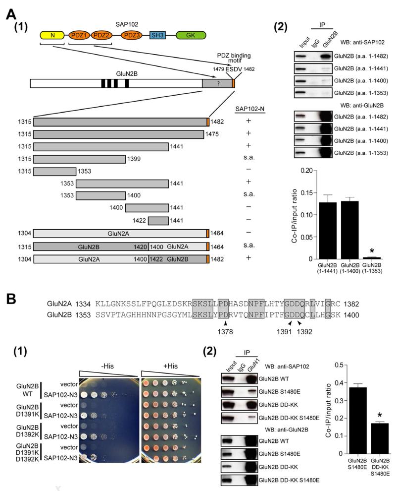
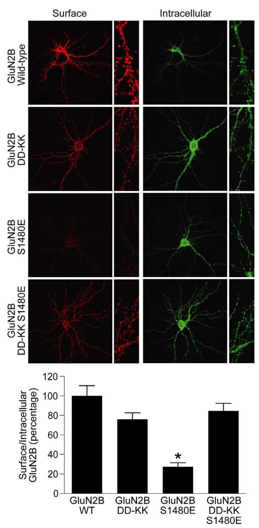
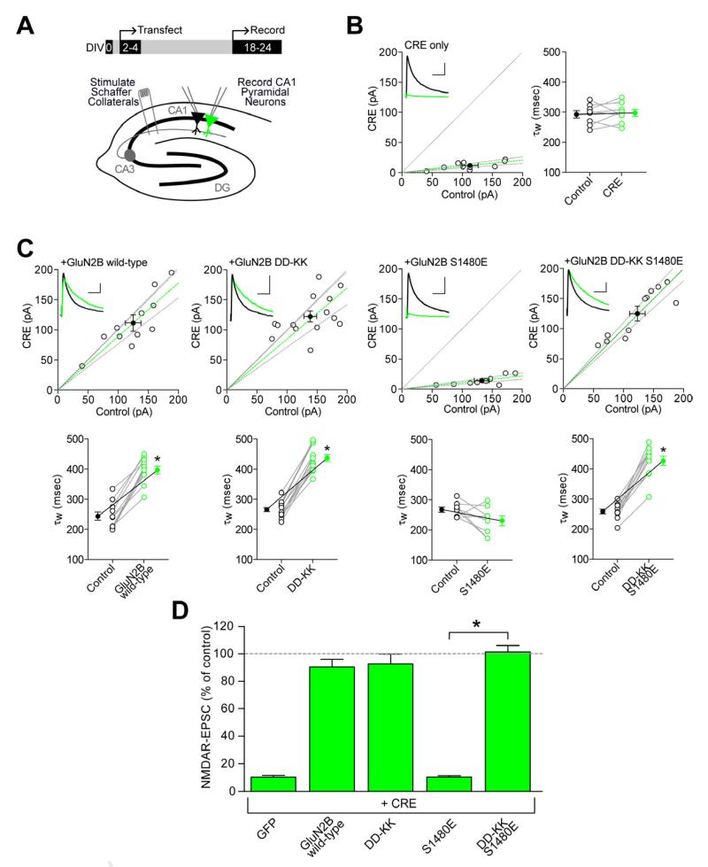
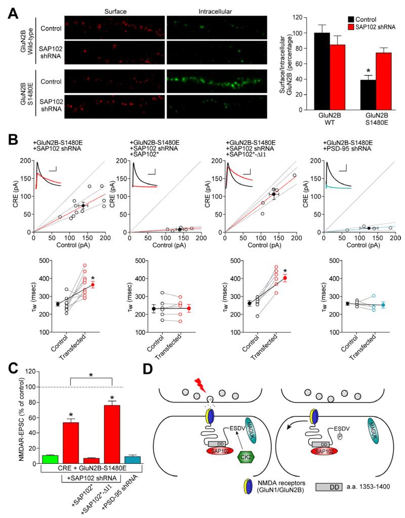
Membrane-associated guanylate kinases (MAGUKs) are the major family of scaffolding proteins at the postsynaptic density. The PSD-MAGUK subfamily, which includes PSD-95, PSD-93, SAP97, and SAP102, is well accepted to be primarily involved in the synaptic anchoring of numerous proteins, including N-methyl-D-aspartate receptors (NMDARs). Notably, the synaptic targeting of NMDARs depends on the binding of the PDZ ligand on the GluN2B subunit to MAGUK PDZ domains, as disruption of this interaction dramatically decreases NMDAR surface and synaptic expression. We recently reported a secondary interaction between SAP102 and GluN2B, in addition to the PDZ interaction. Here, we identify two critical residues on GluN2B responsible for the non-PDZ binding to SAP102. Strikingly, either mutation of these critical residues or knockdown of endogenous SAP102 can rescue the defective surface expression and synaptic localization of PDZ binding-deficient GluN2B. These data reveal an unexpected, nonscaffolding role for SAP102 in the synaptic clearance of GluN2B-containing NMDARs.


Figures

Figure 1
Figure 1. Two critical residues in GluN2B regulate the PDZ-independent interaction with SAP102
(A) (1) Schematics of PDZ-dependent and PDZ-independent interactions between GluN2B and SAP102. Various constructs are shown aligned under full-length GluN2B. The interaction of GluN2B constructs with the N-terminal domain of SAP102 are shown, as measured by the yeast two-hybrid binding assay. s.a. indicates there was self activation of the expression construct so the interaction assay could not be performed. (2) HEK-293 cells were transfected with GFP-GluN2B (a.a. 1-1482), GFP-GluN2B (a.a. 1-1441), GFP-GluN2B (a.a. 1-1400) or GFP-GluN2B (a.a. 1-1353) and SAP102. Receptors were immunoprecipitated from cell lysates with anti-GluN2B antibodies or IgG antibodies as a negative control. Immunoprecipitates were resolved by SDS-PAGE and immunoblotted with anti-SAP102 or anti-GluN2B antibodies. Input = 10 % of total cell lysate. The data were quantified by measuring co-IP/input SAP102 band intensity ratios using ImageJ software. Data represent means ± SEM (N = 3 independent experiments). See also Figure S1. (B) An alignment of GluN2A (1334-1382) and GluN2B (1353-1400) are shown. GluN2B D1378, D1391 and D1392 are indicated with arrowheads. The N3 domain of SAP102 (101-148) is the minimum region required for the non-PDZ interaction (Chen et al., 2011) (1) Yeast were co-transformed with LexA-GluN2B, LexA-GluN2B D1391K, LexA-GluN2B D1392K, or LexA-GluN2B D1391K D1392K and Gal4 vector or Gal4-SAP102-N3, and growth was evaluated on appropriate yeast selection medium. Results shown are 10-fold serial dilutions of yeast cells. (2) HEK-293 cells were transfected with GluN1, SAP102 and GluN2B constructs. Receptors were immunoprecipitated from cell lysates with anti-GluN1 antibodies or IgG antibodies as a negative control. Immunoprecipitates were immunoblotted with anti-SAP102 or anti-GluN2B antibodies. Input = 5 % of total cell lysate. The data were quantified by measuring Co-IP/input SAP102 band intensity ratios using ImageJ software. Data represent means ± SEM (N = 3 independent experiments; *p<0.01). See also Figure S1.
Figure 2
Figure 2. DD-KK mutations rescue the surface expression of GluN2B S1480E in neurons
Hippocampal neurons were transfected with GluN2B constructs containing an extracellular GFP tag. Surface staining was performed with anti-GFP and Alexa 568-conjugated (red) anti-rabbit secondary antibody, followed by fixation and permeabilization, and the internal pool of receptors was labeled with anti-GFP and Alexa 488-conjugated (green) anti-rabbit secondary antibody. Data represent means ± SEM. (n=27; *p<0.01) (N = 3 independent experiments). See also Figure S2.
Figure 3
Figure 3. Disruption of non-PDZ SAP102 binding rescues the synaptic targeting of PDZ-deficient GluN2B
(A) Organotypic hippocampal slice cultures were made from P7 Grin2afl/flGrin2bfl/fl mice, biolistically transfected on DIV2-4, and paired whole-cell recordings were obtained from Cre-expressing and neighboring CA1 pyramidal neurons on DIV18-24. (B-C) Scatter plots of peak amplitudes of NMDAR-EPSCs from single pairs (open circles) and mean ± SEM (filled circles) from transfected and control cells. Dashed lines represent linear regression and 95% confidence interval. Sample traces are as follows: control cell, black; transfected cell, green; scale bars represent 100 msec and 40 pA. NMDAR-EPSC decay times expressed in msec as a weighted tau (τw) from paired transfected and control cells. (B) Transfection with Cre alone. (C) Co-transfection of Cre with wild-type GluN2B, GluN2B DD-KK, GluN2B S1480E or the double GluN2B mutant, DD-KK S1480E. Decay kinetics were analyzed by a paired Student’s t-test, *p<0.0001. (D) Summary graph of data. Bars represent the mean ± SEM of the ratios of transfected to control cells from each pair, expressed as percentages. Data were analyzed by the Mann-Whitney U test, *p<0.0001 compared with GluN2B-S1480E. Actual values for all data can be found in Supplemental Table S1.
Figure 4
Figure 4. SAP102 controls GluN2B synaptic expression
(A) SAP102 was knocked-down in hippocampal cultures by the lentiviral induction of a specific shRNA at DIV5 and Flag-tagged GluN2B constructs (wt or S1480E) were transfected at DIV10. Surface staining was performed with anti-Flag and Alexa-568 secondary antibodies (red) and, after permeabilization, and the intracellular pool was labeled with anti-Flag and Alexa-663 secondary antibodies (green). n for GluN2B wt (+/− shRNA) = 26, 26; n for S1480E (+/− shRNA)= 35, 32 (N = 4 independent experiments). Data represent means ± SEM. *p<0.001. See also Figure S3. (B) Co-transfection of Cre with GluN2B S1480E, shRNA against mouse SAP102, shRNA-proof SAP102 variants (SAP102*), or shRNA against mouse PSD-95 in hippocampal slice cultures from P7 Grin2afl/flGrin2bfl/fl mice. Top, scatter plot of peak amplitudes of NMDAR-EPSCs from single pairs (open circles) and mean ± SEM (filled circles) from transfected and control cells. Dashed lines represent linear regression and 95% confidence interval. Sample traces are as follows: control cell, black; transfected cell, red; scale bars represent 100 msec and 40 pA. Bottom, NMDAR-EPSC decay times expressed as a weighted tau (τw) from paired transfected and control cells. Decay kinetics were analyzed by a paired Student’s t-test. See also Figure S3. (C) Bar graph represents mean ± SEM of the ratios of transfected to control cells from each pair, expressed as percentages (GluN2B S1480E is from Figure 3). Data were analyzed by the Mann-Whitney U test, *p<0.0001 compared with GluN2B-S1480E or as indicated. Actual values for all data can be found in Supplemental Table S1. (D) Model for the role of SAP102 in regulating GluN2B-containing NMDARs at the synapse. GluN2B-containing NMDARs are stabilized on synaptic membranes through the PDZ interactions with PSD-MAGUKs (PSD-95, PSD-93, SAP97 or SAP102) and non-PDZ interactions with SAP102. Synaptic activity induces GluN2B phosphorylation on S1480 by CK2, which disrupts the PDZ interaction between synaptic NMDARs and MAGUKS at the post-synaptic density. The secondary interaction of GluN2B with the N-terminus of SAP102 then facilitates the lateral diffusion of NMDARs to perisynaptic endocytic zones.

References

    1. Al-Hallaq RA, Conrads TP, Veenstra TD, Wenthold RJ. NMDA di-heteromeric receptor populations and associated proteins in rat hippocampus. J Neurosci. 2007;27:8334–8343. - PMC - PubMed
    1. Barria A, Malinow R. Subunit-specific NMDA receptor trafficking to synapses. Neuron. 2002;35:345–353. - PubMed
    1. Bellone C, Nicoll RA. Rapid bidirectional switching of synaptic NMDA receptors. Neuron. 2007;55:779–785. - PubMed
    1. Chen BS, Roche KW. Regulation of NMDA receptors by phosphorylation. Neuropharmacology. 2007;53:362–368. - PMC - PubMed
    1. Chen BS, Thomas EV, Sanz-Clemente A, Roche KW. NMDA receptor-dependent regulation of dendritic spine morphology by SAP102 splice variants. J Neurosci. 2011;31:89–96. - PMC - PubMed

Publication types

MeSH terms