. 2012 Nov 29;2(5):1104-10.
doi: 10.1016/j.celrep.2012.09.021. Epub 2012 Oct 25.

The transporter Spns2 is required for secretion of lymph but not plasma sphingosine-1-phosphate

Affiliations

The transporter Spns2 is required for secretion of lymph but not plasma sphingosine-1-phosphate

Alejandra Mendoza et al. Cell Rep. .

Abstract

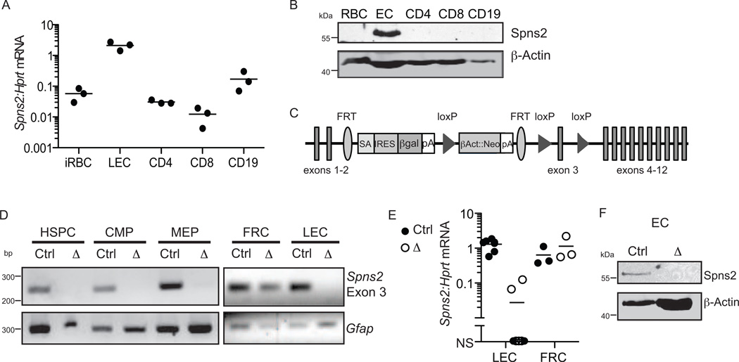
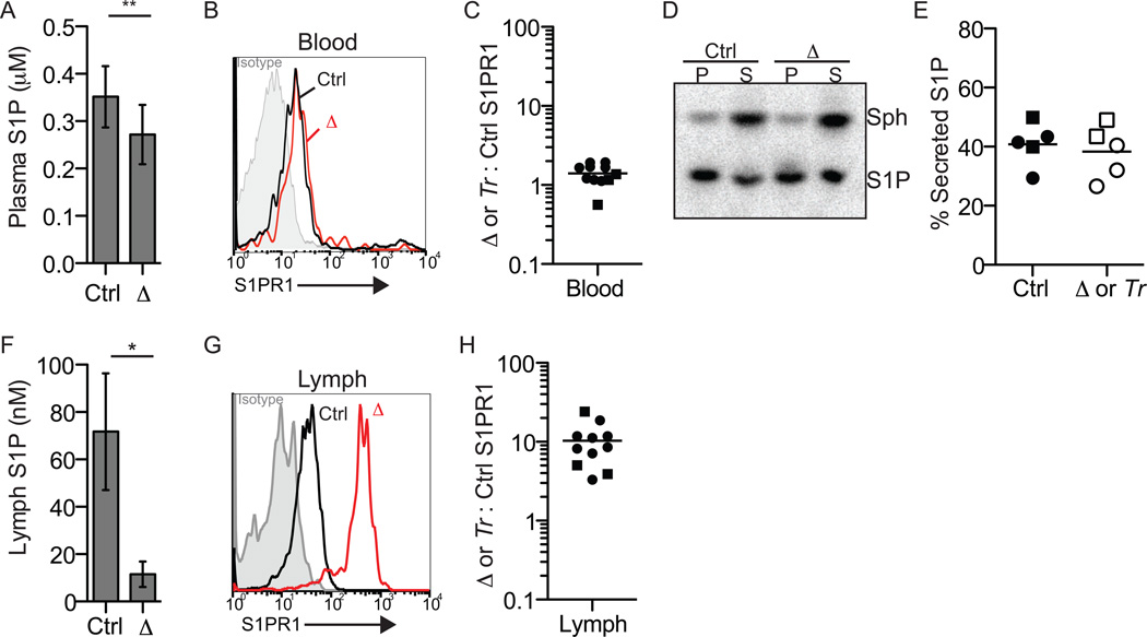
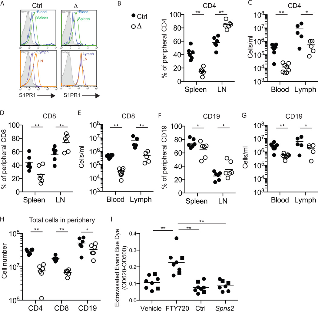
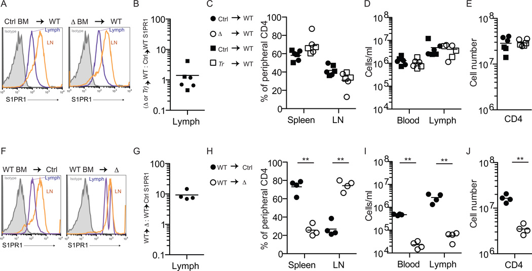
Plasma sphingosine-1-phosphate (S1P) regulates vascular permeability, and plasma and lymph S1P guide lymphocyte egress from lymphoid organs. S1P is made intracellularly, and little is known about how S1P is delivered into circulatory fluids. Here, we find that mice without the major facilitator superfamily transporter Spns2 have a profound reduction in lymph S1P, but only a minor decrease in plasma S1P. Spns2-deficient mice have a redistribution of lymphocytes from the spleen to lymph nodes and a loss of circulating lymphocytes, consistent with normal egress from the spleen directed by plasma S1P and blocked egress from lymph nodes directed by lymph S1P. Spns2 is needed in endothelial cells to supply lymph S1P and support lymphocyte circulation. As a differential requirement for lymph and blood S1P, Spns2 may be an attractive target for immune suppressive drugs.


Figures

Figure 1
Figure 1. Murine Spns2 is expressed by endothelial cells but not RBC
(A) Expression of Spns2 mRNA by the indicated cell populations, expressed relative to hypoxanthine-guanine phosphoribosyltransferase (Hprt) transcript, assessed by RT-qPCR. Ter119+CD71+ immature RBC (iRBC) were sorted from bone marrow; CD31+gp38+ lymphatic endothelial cells (LEC), CD4+CD62Lhi T cells (CD4), CD8+CD62Lhi T cells (CD8), and CD19+CD62Lhi B cells (CD19) were sorted from lymph nodes. Data compile 3 experiments with mice on a B6 background. (B) Expression of Spns2 protein by the indicated cell populations, assessed by Western blot. RBC were isolated from blood by differential centrifugation; CD31+ endothelial cells (EC) were isolated from heart and lung by magnetic bead enrichment; and CD4+ T cells, CD8+ T cells, and CD19+ B cells were isolated from lymph nodes by magnetic bead enrichment. Data are representative of 3 experiments with mice on a B6 background. (C) Spns2-targeted allele. SA: splice acceptor; pA: polyadenylation signal. (D-F) Efficiency of Spns2 deletion in Spns2f/fTie2-Cre+ mice. (D) PCR for Spns2 exon 3 in genomic DNA from sorted Spns2f/fTie2Cre+(Δ) or littermate control (Ctrl) cells. Littermate controls maintained one or two intact alleles of Spns2. RBC progenitors were isolated from bone marrow. Hematopoietic stem and progenitor cells (HSPC) were defined as LinIL7Rαc-Kit+Sca1+; common myeloid progenitors (CMP) as LinIL7Rαc Kit+Sca1FcγRloCD34+; and megakaryocyte erythroid progenitors (MEP) as LinIL7Rαc-Kit+Sca1FcγRloCD34. Lymphatic endothelial cells (LEC), defined as CD45CD31+gp38+, and fibroblastic reticular cells (FRC), defined as CD45CD31gp38+ were isolated from lymph nodes. Data are representative of at least 2 experiments. (E) Spns2 mRNA assessed by RT-qPCR of transcripts from sorted LEC and FRC. Data compile 7 pairs of mice analyzed in 7 experiments for LEC and 3 pairs of mice analyzed in 3 experiments for FRC. NS, no signal. (F) Spns2 protein assessed by Western blot of mixed heart and lung endothelial cells (EC). Data are representative of 2 experiments. See also Fig. S1.
Figure 2
Figure 2. Spns2 is essential to supply lymph S1P, but makes a minor contribution to plasma S1P
(A-E) Spns2 makes a minor contribution to plasma S1P. (A) Plasma S1P of Spns2f/fTie2-Cre+ (Δ) and littermate control mice quantified by mass spectrometry (n=6, error bars show standard deviation). (B) Surface S1PR1 on CD62LhiCD4+T cells circulating in the blood of a representative Spns2f/fTie2-Cre+ mouse (red) and its littermate control (black). Isotype control is shaded grey; note that based on staining of S1PR1 knockout animals, the isotype control staining may be artificially low (Green et al. 2011 and data not shown). (C) The ratio of surface S1PR1 MFI on CD62LhiCD4+T cells in the blood of a Spns2f/fTie2-Cre+ or Spns2tr/tr (Tr) mouse to surface S1PR1 MFI on CD62LhiCD4+ T cells in the blood of its littermate control. Circles indicate Spns2f/fTie2-Cre+ mice and controls (8 pairs analyzed in 7 experiments), and squares indicate Spns2tr/tr mice and controls (3 pairs analyzed in 3 experiments). (D-E) Spns2f/fTie2-Cre+ or Spns2tr/tr and littermate control RBC were incubated with [3-3H]sphingosine (Sph), which crosses the plasma membrane into the cytosol where it can be phosphorylated. After 90 minutes, the cell pellet (P) and supernatant (S) were collected. Extracted lipids were separated by thin layer chromatography (TLC) to assess the distribution of [3-3H]S1P. (D) A representative TLC plate visualized by Phosphorimager. (E) Data pooled from 5 experiments. % secreted S1P: 100×[S1P in supernatant]/[S1P in pellet + S1P in supernatant]. Circles indicate Spns2f/fTie2-Cre+ mice and controls, and squares indicate Spns2tr/tr mice and controls. (F-H) Spns2 is essential to supply lymph S1P. (F) Lymph S1P of Spns2f/fTie2-Cre+ mice and littermate controls quantified by mass spectrometry (n=2-3, error bars show standard deviation). (G) Surface S1PR1 on CD62LhiCD4+ T cells circulating in the lymph of a representative Spns2f/fTie2-Cre+ mouse (red) and its littermate control (black). Isotype control is shaded grey. (H) The ratio of surface S1PR1 MFI on CD62LhiCD4+ T cells in the lymph of a Spns2f/fTie2-Cre+ or Spns2tr/tr (Tr) mouse to surface S1PR1 MFI on CD62LhiCD4+ T cells in the lymph of its littermate control. Circles indicate Spns2f/fTie2-Cre+ mice and controls (8 pairs analyzed in 7 experiments), and squares indicate Spns2tr/tr mice and controls (3 pairs analyzed in 3 experiments). *p<0.05, **p<0.01
Figure 3
Figure 3. Spns2 is required for peripheral lymphocyte circulation
(A) Surface S1PR1 on CD62LhiCD4+ T cells in the blood and spleen (top panels) and in the lymph and lymph nodes (bottom panels) of a representative Spns2f/fTie2Cre+ mouse (Δ) and its littermate control (Ctrl). Isotype control is shaded grey. Data are representative of 8 pairs of mice analyzed in 7 experiments. (B-G) Lymphocyte distribution in Spns2f/fTie2Cre+ mice and littermate controls. (B,D,F) Percent of total peripheral CD62LhiCD4+ T cells (B), CD62LhiCD8+ T cells (D), and CD62LhiCD19+ B cells (F) in the spleen and lymph nodes (LN). Total peripheral lymphocytes are defined as those in spleen and a subset of LN (brachial, axillary, inguinal, and mesenteric); blood and lymph make a negligible contribution. [For example, (B, spleen) shows 100×(# CD62LhiCD4+ T cells in spleen)/(# CD62LhiCD4+ T cells in spleen + # CD62LhiCD4+ T cells in LN).] Data pool 6 pairs of mice analyzed in 5 experiments. (C,E,G) Total number of CD62LhiCD4+ T cells (C), CD62LhiCD8+ T cells (E), and CD62LhiCD19+ B cells (G) in blood and lymph. Data pool 8 pairs of mice analyzed in 7 experiments for blood, and 5 pairs of mice analyzed in 4 experiments for lymph. (H) Total number of CD62LhiCD4+ T cells, CD62LhiCD8+ T cells, and CD62LhiCD19+ B cells in the periphery. Data pool 6 pairs of mice analyzed in 5 experiments. (I) Vascular permeability in Spns2f/fTie2Cre+ mice and littermate controls. Spns2-deficient mice and littermate controls, and C57BL6 mice treated with FTY720 or vehicle, were injected intravenously with Evans Blue dye. After 90 minutes, mice were perfused with PBS. Lungs were removed and extravasated Evans Blue dye was quantified by spectrophotometry. Circles indicate Spns2f/fTie2-Cre+ mice and controls (6 groups analyzed in 6 experiments), and squares indicate Spns2tr/tr mice and controls (2 groups analyzed in 2 experiments). *p<0.05, **p<0.01. See also Fig. S2.
Figure 4
Figure 4. Spns2 is required in endothelial cells to supply lymph S1P and support lymphocyte circulation
(A-E) Ubiquitin C:GFP+ mice were lethally irradiated and reconstituted with BM from Spns2-deficient mice or littermate controls (GFP+ mice were used to allow assessment of RBC chimerism). Mice were analyzed >6 weeks after transplantation, when RBC and circulating lymphocytes were 97–99% donor-derived. Deletion of Spns2 in Spns2f/fTie2Cre+ (Δ) donor hematopoietic stem and progenitor cells (HSPC) was confirmed by PCR. Data compile 6 pairs of recipients, with 2 pairs of Spns2f/fTie2-Cre+/control BM donors (circles) and 1 pair of Spns2tr/tr (Tr)/control BM donors (squares), analyzed in 6 experiments. (A) Representative histogram of surface S1PR1 on CD62LhiCD4+ T cells in the lymph and lymph nodes of the indicated chimeras. Isotype control is shaded grey. (B) The ratio of surface S1PR1 MFI on CD62LhiCD4+ T cells in the lymph of a WT mouse with Spns2-deficient BM to surface S1PR1 MFI on CD62LhiCD4+ T cells in the lymph of a WT mouse with littermate control BM. (C) Percent of total donor-derived peripheral CD62LhiCD4+ T cells in the spleen and lymph nodes. (D) Total number of donor-derived CD62LhiCD4+ T cells in blood and lymph. (E) Total number of donor-derived CD62LhiCD4+ T cells in the periphery. (F-J) Spns2f/fTie2-Cre+ mice and littermate controls were lethally irradiated and reconstituted with BM from Ubiquitin C:GFP+ mice. Mice were analyzed >6 weeks after transplantation, when RBC were 88–99% donor-derived and lymphocytes were 85–98% donor-derived. Data compile 4 pairs of mice analyzed in 4 experiments. (F) Representative histogram of surface S1PR1 on CD62LhiCD4+ T cells in the lymph and lymph nodes of the indicated chimeras. Isotype control is shaded grey. (G) The ratio of surface S1PR1 MFI on CD62LhiCD4+ T cells in the lymph of a Spns2f/fTie2-Cre+ mouse with WT BM to surface S1PR1 MFI on CD62LhiCD4+ T cells in the lymph of its littermate control with WT BM. (H) Percent of total donor-derived peripheral CD62LhiCD4+ T cells in the spleen and lymph nodes. (I) Total number of donor-derived CD62LhiCD4+ T cells in blood and lymph. (J) Total number of donor-derived CD62LhiCD4+ T cells in the periphery. *p<0.05, **p<0.01. See also Fig. S3 and Fig. S4.

References

    1. Breart B, Ramos-Perez WD, Mendoza A, Salous AK, Gobert M, Huang Y, Adams RH, Lafaille JJ, Escalante-Alcalde D, Morris AJ, et al. Lipid phosphate phosphatase 3 enables efficient thymic egress. J Exp Med. 2011;208:1267–1278. - PMC - PubMed
    1. Brinkmann V, Billich A, Baumruker T, Heining P, Schmouder R, Francis G, Aradhye S, Burtin P. Fingolimod (FTY720): discovery and development of an oral drug to treat multiple sclerosis. Nat Rev Drug Discov. 2010;9:883–897. - PubMed
    1. Camerer E, Regard JB, Cornelissen I, Srinivasan Y, Duong DN, Palmer D, Pham TH, Wong JS, Pappu R, Coughlin SR. Sphingosine-1-phosphate in the plasma compartment regulates basal and inflammation-induced vascular leak in mice. J Clin Invest. 2009;119:1871–1879. - PMC - PubMed
    1. Ficara F, Murphy MJ, Lin M, Cleary ML. Pbx1 regulates self-renewal of long-term hematopoietic stem cells by maintaining their quiescence. Cell stem cell. 2008;2:484–496. - PMC - PubMed
    1. Fukuhara S, Simmons S, Kawamura S, Inoue A, Orba Y, Tokudome T, Sunden Y, Arai Y, Moriwaki K, Ishida J, et al. The sphingosine-1-phosphate transporter Spns2 expressed on endothelial cells regulates lymphocyte trafficking in mice. J Clin Invest. 2012;122:1416–1426. - PMC - PubMed

Publication types