

Telmisartan attenuates hepatic fibrosis in bile duct-ligated rats
- PMID: 23103625
- PMCID: PMC4001837
- DOI: 10.1038/aps.2012.115
Telmisartan attenuates hepatic fibrosis in bile duct-ligated rats
Abstract
Aim: To evaluate the antifibrotic effect of telmisartan, an angiotensin II receptor blocker, in bile duct-ligated rats.
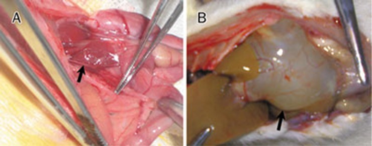
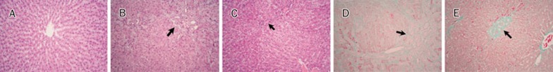
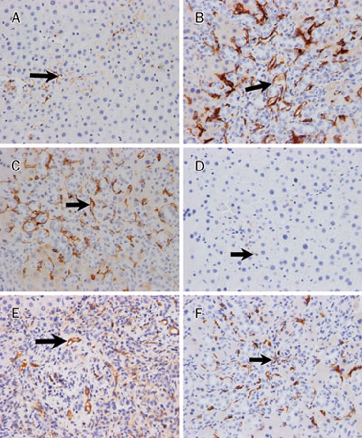
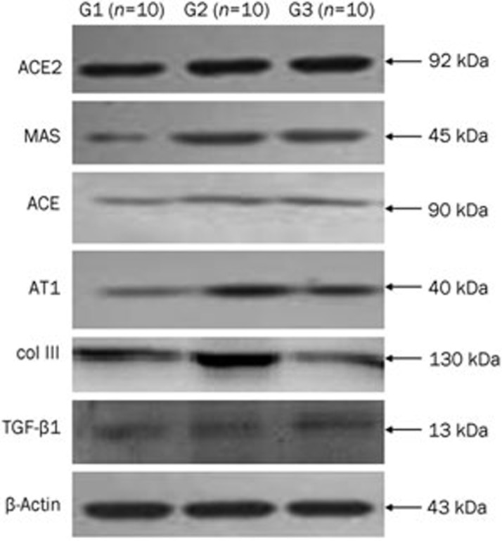
Methods: Adult Sprague-Dawley rats were allocated to 3 groups: sham-operated rats, model rats underwent common bile duct ligation (BDL), and BDL rats treated with telmisartan (8 mg/kg, po, for 4 weeks). The animals were sacrificed on d 29, and liver histology was examined, the Knodell and Ishak scores were assigned, and the expression of angiotensin-converting enzyme (ACE) and ACE2 was evaluated with immunohistochemical staining. The mRNAs and proteins associated with liver fibrosis were evaluated using RTQ-PCR and Western blot, respectively.
Results: The mean fibrosis score of BDL rats treated with telmisartan was significantly lower than that of the model rats (1.66±0.87 vs 2.13±0.35, P=0.015). However, there was no significant difference in inflammation between the two groups, both of which showed moderate inflammation. Histologically, treatment with telmisartan significantly ameliorated BDL-caused the hepatic fibrosis. Treatment with telmisartan significantly upregulated the mRNA levels of ACE2 and MAS, and decreased the mRNA levels of ACE, angiotensin II type 1 receptor (AT1-R), collagen type III, and transforming growth factor β1 (TGF-β1). Moreover, treatment with telmisartan significantly increased the expression levels of ACE2 and MAS proteins, and inhibited the expression levels of ACE and AT1-R protein.
Conclusion: Telmisartan attenuates liver fibrosis in bile duct-ligated rats via increasing ACE2 expression level.
Figures

Similar articles
-
Angiotensin receptor blockers are superior to angiotensin-converting enzyme inhibitors in the suppression of hepatic fibrosis in a bile duct-ligated rat model.J Gastroenterol. 2008;43(11):889-96. doi: 10.1007/s00535-008-2239-9. Epub 2008 Nov 18. J Gastroenterol. 2008. PMID: 19012043
-
Effects of Yinchenhao decoction on self-regulation of renin-angiotensin system by targeting angiotensin converting enzyme 2 in bile duct-ligated rat liver.J Huazhong Univ Sci Technolog Med Sci. 2015 Aug;35(4):519-524. doi: 10.1007/s11596-015-1463-9. Epub 2015 Jul 31. J Huazhong Univ Sci Technolog Med Sci. 2015. PMID: 26223920
-
Cardioprotective effects of telmisartan against heart failure in rats induced by experimental autoimmune myocarditis through the modulation of angiotensin-converting enzyme-2/angiotensin 1-7/mas receptor axis.Int J Biol Sci. 2011;7(8):1077-92. doi: 10.7150/ijbs.7.1077. Epub 2011 Sep 8. Int J Biol Sci. 2011. PMID: 21927577 Free PMC article.
-
[Inhibitory effect of angiotensin blockade on hepatic fibrosis in common bile duct-ligated rats].Korean J Hepatol. 2007 Mar;13(1):61-9. Korean J Hepatol. 2007. PMID: 17380076 Korean.
-
Telmisartan, an AT1 receptor blocker and a PPAR gamma activator, alleviates liver fibrosis induced experimentally by Schistosoma mansoni infection.Parasit Vectors. 2013 Jul 5;6:199. doi: 10.1186/1756-3305-6-199. Parasit Vectors. 2013. PMID: 23829789 Free PMC article.
Cited by
-
Radiation Induced Skin Fibrosis (RISF): Opportunity for Angiotensin II-Dependent Intervention.Int J Mol Sci. 2024 Jul 29;25(15):8261. doi: 10.3390/ijms25158261. Int J Mol Sci. 2024. PMID: 39125831 Free PMC article. Review.
-
Olmesartan Improves Hepatic Sinusoidal Remodeling in Mice with Carbon Tetrachloride-Induced Liver Fibrosis.Biomed Res Int. 2022 Aug 26;2022:4710993. doi: 10.1155/2022/4710993. eCollection 2022. Biomed Res Int. 2022. PMID: 36060127 Free PMC article.
-
A comprehensive guide to the pharmacologic regulation of angiotensin converting enzyme 2 (ACE2), the SARS-CoV-2 entry receptor.Pharmacol Ther. 2021 May;221:107750. doi: 10.1016/j.pharmthera.2020.107750. Epub 2020 Dec 1. Pharmacol Ther. 2021. PMID: 33275999 Free PMC article. Review.
-
Stimulation of the Angiotensin II AT2 Receptor is Anti-inflammatory in Human Lipopolysaccharide-Activated Monocytic Cells.Inflammation. 2015 Aug;38(4):1690-9. doi: 10.1007/s10753-015-0146-9. Inflammation. 2015. PMID: 25758542
-
The Renin Angiotensin System as a Therapeutic Target in Traumatic Brain Injury.Neurotherapeutics. 2023 Oct;20(6):1565-1591. doi: 10.1007/s13311-023-01435-8. Epub 2023 Sep 27. Neurotherapeutics. 2023. PMID: 37759139 Free PMC article. Review.
References
-
- Pereira RM, Dos Santos RA, Teixeira MM, Leite VH, Costa LP, da Costa Dias FL, et al. The renin-angiotensin system in a rat model of hepatic fibrosis: evidence for a protective role of angiotensin-(1–7) J Hepatol. 2007;46:674–81. - PubMed
-
- Castro-Chaves P, Cerqueira R, Pintalhao M, Leite-Moreira AF. New pathways of the renin-angiotensin system: the role of ACE2 in cardiovascular pathophysiology and therapy. Expert Opin Ther Targets. 2010;14:485–96. - PubMed
-
- Dejima T, Tamura K, Wakui H, Maeda A, Ohsawa M, Kanaoka T, et al. Prepubertal angiotensin blockade exerts long-term therapeutic effect through sustained ATRAP activation in salt-sensitive hypertensive rats. J Hypertens. 2011;29:1919–29. - PubMed
Publication types
MeSH terms
Substances
LinkOut - more resources
Full Text Sources
Other Literature Sources
Medical
Research Materials
Miscellaneous
