Review
. 2013 Mar;1831(3):555-74.
doi: 10.1016/j.bbalip.2012.10.006. Epub 2012 Oct 26.

Mammalian P4-ATPases and ABC transporters and their role in phospholipid transport

Affiliations
Review

Mammalian P4-ATPases and ABC transporters and their role in phospholipid transport

Jonathan A Coleman et al. Biochim Biophys Acta. 2013 Mar.

Abstract

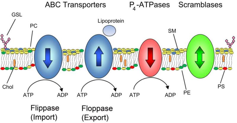
Transport of phospholipids across cell membranes plays a key role in a wide variety of biological processes. These include membrane biosynthesis, generation and maintenance of membrane asymmetry, cell and organelle shape determination, phagocytosis, vesicle trafficking, blood coagulation, lipid homeostasis, regulation of membrane protein function, apoptosis, etc. P(4)-ATPases and ATP binding cassette (ABC) transporters are the two principal classes of membrane proteins that actively transport phospholipids across cellular membranes. P(4)-ATPases utilize the energy from ATP hydrolysis to flip aminophospholipids from the exocytoplasmic (extracellular/lumen) to the cytoplasmic leaflet of cell membranes generating membrane lipid asymmetry and lipid imbalance which can induce membrane curvature. Many ABC transporters play crucial roles in lipid homeostasis by actively transporting phospholipids from the cytoplasmic to the exocytoplasmic leaflet of cell membranes or exporting phospholipids to protein acceptors or micelles. Recent studies indicate that some ABC proteins can also transport phospholipids in the opposite direction. The importance of P(4)-ATPases and ABC transporters is evident from the findings that mutations in many of these transporters are responsible for severe human genetic diseases linked to defective phospholipid transport. This article is part of a Special Issue entitled Phospholipids and Phospholipid Metabolism.


Figures

Fig. 1
Fig. 1
Regulation of lipid asymmetry in biological membranes. The distribution of lipids in biological membranes is regulated by three distinct families of membrane transporters: ABC transporters, P4-ATPases, and scramblases. Most ABC transporters catalyze the ATP-dependent transport of lipids from the cytoplasmic leaflet of the bilayer to the extracellular (lumenal) side of the membrane while P4-ATPases transport in the opposite direction. ABC transporters can efflux lipids to lipoproteins such as Apo-A1 in the case of ABCA1 or to bile micelles. Recent studies indicate that a few ABC transporters are importers or flippases transporting phospholipids in the same direction as P4-ATPases. Scramblases are energy independent transporters and act to abolish lipid asymmetry by randomizing lipid distributions. Lipids such as PC, SM, and glycolipids are found in the extracellular or lumenal leaflet while the aminophospholipids PE and PS are preferentially on the cytosolic leaflet. Abbreviations used: PC, phosphatidylcholine; SM, sphingomyelin; PE, phosphatidylethanolamine; PS, phosphatidylserine; GSL, glycosphingolipid; Chol, cholesterol.
Fig. 2
Fig. 2
Phylogenetic tree of human P-type and P4-ATPases. (A) There are 36 different genes which encode for P-type ATPases in humans. Type IB are heavy metal pumps, Type IIA and Type IIB are Ca2+ pumps, and Type IIC are Na+/K+ or H+/K+ pumps. Type IV ATPases transport phospholipids. Type V ATPases have no assigned specificity. Type IA and Type III pumps are not found in humans. (B) There are 14 different Type IV ATPases in humans which are organized into five different subfamilies according to sequence. Trees were generated using ClustalX and visualized using the Interactive Tree of Life online tool (http://itol.embl.de/).
Fig. 3
Fig. 3
Domain structure, organization, and proposed mechanism of P4-ATPases. (A) P4-ATPases adopt a four domain structure consisting of cytosolic A (actuator), P (phosphorylation), and N (nucleotide binding) domains as well as a M (membrane) domain consisting of 10 membrane spanning segments and containing the translocation pathway. Absolutely conserved motifs are shown. P-type ATPases are phosphorylated at the aspartic acid of the invariant DKTG motif. The glutamic acid of the DGE motif in the A domain catalyzes dephosphorylation. The CDC50 β-subunit is shown containing four N-linked glycosylations and two disulfide linkages in the large E (extracellular) domain. (B) P-type ATPases exist in four primary conformations. Binding of ATP and phosphorylation of the P-domain in the E1 form converts the enzyme to E1P. During the E1P to E2P transition, the A-domain rotates allowing lipids to bind to an extracellular binding site in the M-domain in the E2P conformation. Dephosphorylation of E2P drives the translocation of the lipid through the membrane to the cytoplasmic side. The enzyme is converted back to the E1 form when the A-domain moves away from the P-domain. The role of CDC50 (green) is unknown.
Fig 4
Fig 4
Predicted two-dimensional topology of lipid ABC transporters and models of ABC transporter mechanism. A. The two transmembrane domains (TMDs) and nucleotide binding domains (NBDs) arrange to form full transporter and are represented by some ABCB members and ABCA members with additional exocytosolic domains. Some ABCC family members are full transporters with an additional N-terminal TMD. Half transporters in ABCB and ABCG subfamilies have different modular organizations of a TMD and NBD. B. A simplified‘Switch Model’: In the inward-facing conformation, the NBDs are separated and nucleotide-free. Phospholipid substrate entry into the substrate binding site occurs from the cytosolic leaflet of the membrane bilayer. ATP-dependent dimerization of NBDs progresses to pull the TMDs from an inward- to outward-facing conformation. Phospholipid is translocated to the extracellular side of the membrane and ATP hydrolysis resets the transporter in the inward conformation. C. A simplified ‘Constant-contact Model’: The two NBDs remain associated in the sandwich dimer form throughout the transport cycle. Inward and outward conformations are directed via subunit conformational changes and the twisting motion of NBDs. ATP hydrolysis alternates with one site hydrolyzing nucleotide that facilitates the binding and hydrolysis in the other site.
Fig 5
Fig 5
Apo-A1 lipidation via ABCA1 activity within plasma membrane microdomains. Lipid free apo-A1 initially interacts with monomeric or oligomeric ABCA1. The PC flippase activity of ABCA1 creates a High Capacity Binding Site (HCBS) within cholesterol poor microdomains. Phospholipid translocation via ABCA1 may also induce membrane bending of the bilayer to create an exovesiculated binding site. ABCA1 subsequently mediates transfer of apo-A1 to these HCBS populations, from which apo-A1 selectively microsolubilizes PC to become discoidal apo-A1 species. Further microsolubilization of cholesterol and PC generates nascent HDL with two, three, and four molecules of apo-A1.
Fig 6
Fig 6
A. Overview of ABC transporters involved in lipid efflux. Schematic representation of general ABC transporter localization, known acceptors and direction of transport. Vectorial transport depicted by black arrows at the plasma membrane. Vectorial transport in many ABC transporters and by intracellular ABC transporters has not been firmly established. B. Hepatocytic ABC transporters. Newly synthesized bile salts are effluxed by ABCB11, where they form micelles with PC translocated to the outer leaflet of the canalicular membrane by ABCB4. ABCG5/G8 translocates cholesterol into the bile lipid mixture.The action of PS translocation to the inner leaflet by ATP8B1 maintains the lipid asymmetry at the canalicular membrane. C. Role of ABCA4 in photoreceptor disc membranes. Photoexcitation of disc membranes in photoreceptors causes release of all-trans retinal from rhodopsin. Retinal is reduced in the visual cycle or reacts with PE in disc membranes to form N-ret PE. ABCA4 binds and translocates N-ret PE from the lumen to the cytosolic side of the disc membrane. All-trans retinal then dissociates from N-ret PE and retinal is shuttled into the visual cycle. ABCA4 also translocates PE from the lumen to the cytosolic side of the disc membrane. D. Surfactant production in alveolus. In alveolar type II cells, ABCA3 present in lamellar bodies translocates PG, PC, and PE which is then secreted into the epithelial lining of the surfactant. E. Epidermal production in skin. ABCA12 transports GlcCer and Cer into the lamellar body which is then redistributed into the cell periphery. Apo-A1/E, apolipoprotein; HDL High Density Lipoprotein; LDL, low density lipoproteins; SR-B1, Scavenger receptor Class B1 protein; Chol, cholesterol; PC, phosphatidycholine, PE, phosphatidylethanolamine; PS, phosphatidylserine; Cer, ceramine; GlcCer, glucosylceramide; N-Ret PE, N-retinyldiene PE; SM, sphingomyelin

References

    1. Holthuis JC, Levine TP. Lipid traffic: floppy drives and a superhighway. Nat Rev Mol Cell Biol. 2005;6:209–220. - PubMed
    1. van Meer G, Voelker DR, Feigenson GW. Membrane lipids: where they are and how they behave. Nat Rev Mol Cell Biol. 2008;9:112–124. - PMC - PubMed
    1. Daleke DL. Phospholipid flippases. J Biol Chem. 2007;282:821–825. - PubMed
    1. Pomorski T, Menon AK. Lipid flippases and their biological functions. Cell Mol Life Sci. 2006;63:2908–2921. - PMC - PubMed
    1. Folmer DE, Elferink RP, Paulusma CC. P4 ATPases - lipid flippases and their role in disease. Biochim Biophys Acta. 2009;1791:628–635. - PubMed

Publication types

MeSH terms