. 2012 Dec;44(12):1302-9.
doi: 10.1038/ng.2434. Epub 2012 Oct 28.

Inhibition of RNA lariat debranching enzyme suppresses TDP-43 toxicity in ALS disease models

Affiliations

Inhibition of RNA lariat debranching enzyme suppresses TDP-43 toxicity in ALS disease models

Maria Armakola et al. Nat Genet. 2012 Dec.

Abstract

Amyotrophic lateral sclerosis (ALS) is a devastating neurodegenerative disease primarily affecting motor neurons. Mutations in the gene encoding TDP-43 cause some forms of the disease, and cytoplasmic TDP-43 aggregates accumulate in degenerating neurons of most individuals with ALS. Thus, strategies aimed at targeting the toxicity of cytoplasmic TDP-43 aggregates may be effective. Here, we report results from two genome-wide loss-of-function TDP-43 toxicity suppressor screens in yeast. The strongest suppressor of TDP-43 toxicity was deletion of DBR1, which encodes an RNA lariat debranching enzyme. We show that, in the absence of Dbr1 enzymatic activity, intronic lariats accumulate in the cytoplasm and likely act as decoys to sequester TDP-43, preventing it from interfering with essential cellular RNAs and RNA-binding proteins. Knockdown of Dbr1 in a human neuronal cell line or in primary rat neurons is also sufficient to rescue TDP-43 toxicity. Our findings provide insight into TDP-43-mediated cytotoxicity and suggest that decreasing Dbr1 activity could be a potential therapeutic approach for ALS.


Conflict of interest statement

COMPETING FINANCIAL INTERESTS

A.D.G. is an inventor on patents and patent applications that have been licensed to FoldRx.

Figures

Figure 1
Figure 1
Dbr1 deletion suppresses TDP-43 toxicity in yeast. a) Yeast spotting assays showing that DBR1 deletion (dbr1Δ) suppresses TDP-43 toxicity (WT or ALS-linked Q331K mutant). Fivefold serial dilution of yeast cells spotted onto glucose (TDP-43 “off”) or galactose (TDP-43 “on”). b) Immunoblotting shows that TDP-43 expression is not affected in dbr1Δ cells compared to WT. The effect of toxicity suppression by dbr1Δ was specific to ALS-linked RNA binding proteins TDP-43 and FUS/TLS because α-syn or htt103Q toxicity was not suppressed by dbr1Δ. Indeed, dbr1Δ caused an increase in htt103Q toxicity. c) A schematic showing the function of Dbr1 in debranching lariats following splicing. Whereas introns are rapidly degraded in WT cells, in dbr1Δ cells intronic lariats accumulate at high levels .
Figure 2
Figure 2
Dbr1 knockdown reduces TDP-43 toxicity in a human neuronal cell line. a) Schematic of approach used to test effect of Dbr1 on TDP-43 toxicity in human cells. Lentiviruses encoding a doxycycline-regulated FLAG-tagged mutant TDP-43 (Q331K) construct were transduced into M17 neuroblastoma cells and stable clones isolated. Addition of doxycycline induced expression of TDP-43 Q331K and resulted in cellular toxicity. Dbr1 expression was knocked down using siRNAs and the effect on TDP-43 toxicity assessed. b) Immunoblot showing doxycyline-induced expression of FLAG-tagged TDP-43 Q331K, which migrates slightly slower than the endogenous untagged TDP-43. c) Expression of TDP-43 Q331K resulted in decreased cellular viability in a dose-dependent manner compared to control cells (either stably transduced with an empty vector or untransduced), as assessed by the MTT assay (see Methods). * P < 0.01, ** P < 0.05, *** P < 0.001, repeated measures two-way ANOVA. Error bars are mean ± S.E.M. d) Cells were transfected with an siRNA against Dbr1 or a non-targeting (NT) siRNA. Immunoblot shows effective knockdown of Dbr1 expression by the Dbr1-specific siRNA but not the non-targeting control siRNA. Untransfected (–) or non-targeting siRNA (NT) transfected cells exhibited decreased viability upon induction of TDP-43 Q331K expression, whereas the Dbr1-specific siRNA increased viability. * P < 0.05, repeated measures two-way ANOVA. Error bars are mean ± S.E.M.
Figure 3
Figure 3
Dbr1 knockdown reduces TDP-43 toxicity in primary rodent neurons. a) Automated fluorescence microscopy of primary rodent cortical neurons transfected with mApple (top row) and TDP-43-EGFP (bottom row). With this system, hundreds of individual neurons are imaged at repeated intervals– two such neurons are depicted here. While one of the neurons lives for the entire experiment (arrowhead), the other neuron has died by 144 hrs (arrow). Scale bar: 10 μm. b) Cumulative hazard plot showing the cumulative risk of death for neurons in each cohort as a function of time. Expression of TDP-43-EGFP (light blue curve, n=380 neurons counted) significantly increases the risk of death over that of neurons expressing EGFP alone (light green curve, n=245 neurons counted, HR 2.2). Knockdown of Dbr1 using 30 nM Dbr1 siRNA (sR-Dbr1) in neurons expressing TDP-43-EGFP (dark blue curve, n=300 neurons counted) decreases the risk of death by 19% compared to neurons expressing TDP-43-EGFP that received scrambled siRNA (sR-Sc) (light blue curve, n=380 neurons counted). Data were pooled from 2 independent experiments, and statistical significance determined using Cox proportional hazards analysis. NS, not significant; *** HR 2.23 and p < 1×10⁻⁹; ** HR 1.83 and p < 1×10⁻⁴; * HR 0.81 and p 0.04. c) Using a Kaplan-Meier survival curve to display the same data demonstrates a similar effect on neuron survival. d) Dbr1 knockdown results in increased percentage of neurons containing cytoplasmic TDP-43 compared to control scrambled (Sc) siRNA treated neurons. Error bars are mean ± S.E.M. # p < 0.0005, two-tailed t test.
Figure 4
Figure 4
Dbr1 lariat debranching enzymatic activity is required for TDP-43 toxicity and TDP-43 toxicity suppression is mediated by lariat intron accumulation. a) Immunoblot shows that HA-tagged WT Dbr1 and debranching activity dead point mutants are expressed at similar levels. b) Yeast spotting assays shows TDP-43 toxicity is suppressed in dbr1Δ cells. Expressing WT DBR1 in dbr1Δ cells restored toxicity. Two Dbr1 mutants (D40A or N85A) that lack debranching activity were unable to restore TDP-43 toxicity to dbr1Δ cells. Expressing mouse Dbr1 in dbr1Δ yeast cells restored TDP-43 toxicity, indicating that the function of Dbr1 is conserved from yeast to mammals. c) Yeast spotting assay shows that TDP-43 toxicity is suppressed in dbr1Δ cells but not in three other deletion strains (upf1Δ, upf2Δ, and xrn1Δ), which each accumulate non-specific RNA species owing to defects in the cellular nonsense mediated decay (NMD) pathway. Schematic of yeast cells accumulating no excess RNA species (WT), lariat RNAs (dbr1Δ) or non-specific linear RNAs (upf1Δ, upf2Δ, or xrn1Δ) are shown next to the spotting assays.
Figure 5
Figure 5
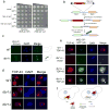
Intronic lariats colocalize with TDP-43 cytoplasmic foci in yeast. a) The toxicity of a TDP-43 mutant (ΔNLS), which is retained in the cytoplasm, is also suppressed by dbr1Δ, suggesting that lariat introns are acting in the cytoplasm to suppress toxicity. b) Strategy to visualize lariat introns in living cells. Homologous recombination was used to insert an MS2 RNA-binding sequence in the intron of the ACT1 gene. A GFP-tagged MS2-CP protein was used to visualize accumulated intronic lariats. c) In WT cells, lariat introns did not accumulate and the MS2-CP-GFP signal was faint and diffusely localized throughout the cell. In contrast, in dbr1Δ cells, MS2-CP-GFP accumulated in one or two bright foci per cell and these were always located in the cytoplasm. Scale bar, 5 μm. d) The size and shape of TDP-43 cytoplasmic inclusions was different in dbr1Δ cells compared to WT. Whereas in WT cells, TDP-43 formed multiple small irregularly-shaped foci (arrowheads), in dbr1Δ cells, there was always at least one large perfectly round focus per cell (arrows) and these always co-localized with lariat introns (see panel e). Scale bar, 5 μm. e) Untagged TDP-43 was expressed in WT and dbr1Δ cells and visualized by immunocytochemistry with a TDP-43-specific antibody. TDP-43 cytoplasmic foci colocalized with tagged lariat introns (arrows). In addition to the alteration in TDP-43 foci size and shape, the size and shape of the lariat introns also appeared to be altered by the presence of TDP-43 (compare MS2-CP-GFP panel in dbr1Δ+vector and dbr1Δ+TDP-43 cells). Scale bar, 5 μm. f) A model of how lariat intron accumulation suppresses TDP-43 cytoplasmic toxicity. In WT cells, TDP-43 aggregates in the cytoplasm and could interfere, via RNA-binding, with essential RNAs and RNA-binding proteins. When Dbr1 activity is inhibited (e.g. in dbr1Δ cells), lariat introns accumulate in the cytoplasm and might act as decoys, sequestering TDP-43 away from interfering with important cellular RNAs.

References

    1. Boillee S, Vande Velde C, Cleveland DW. ALS: a disease of motor neurons and their nonneuronal neighbors. Neuron. 2006;52:39–59. - PubMed
    1. Rosen D, et al. Mutations in Cu/Zn superoxide dismutase gene are associated with familial amyotrophic lateral sclerosis. Nature. 1993;362:59–62. - PubMed
    1. Bruijn LI, et al. Aggregation and motor neuron toxicity of an ALS-linked SOD1 mutant independent from wild-type SOD1. Science. 1998;281:1851–4. - PubMed
    1. Smith RA, et al. Antisense oligonucleotide therapy for neurodegenerative disease. J Clin Invest. 2006;116:2290–6. - PMC - PubMed
    1. Lagier-Tourenne C, Polymenidou M, Cleveland DW. TDP-43 and FUS/TLS: emerging roles in RNA processing and neurodegeneration. Hum Mol Genet. 2010;19:R46–64. - PMC - PubMed

Publication types

Substances