. 2012 Dec;19(12):1363-71.
doi: 10.1038/nsmb.2418. Epub 2012 Oct 28.

Mechanism of repair of 5'-topoisomerase II-DNA adducts by mammalian tyrosyl-DNA phosphodiesterase 2

Affiliations

Mechanism of repair of 5'-topoisomerase II-DNA adducts by mammalian tyrosyl-DNA phosphodiesterase 2

Matthew J Schellenberg et al. Nat Struct Mol Biol. 2012 Dec.

Abstract

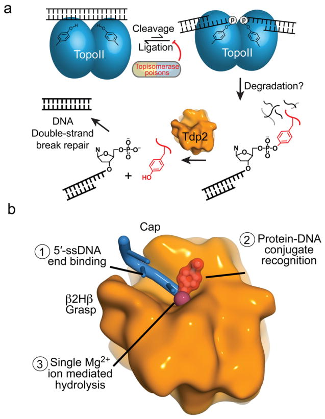
The topoisomerase II (topo II) DNA incision-and-ligation cycle can be poisoned (for example following treatment with cancer chemotherapeutics) to generate cytotoxic DNA double-strand breaks (DSBs) with topo II covalently conjugated to DNA. Tyrosyl-DNA phosphodiesterase 2 (Tdp2) protects genomic integrity by reversing 5'-phosphotyrosyl-linked topo II-DNA adducts. Here, X-ray structures of mouse Tdp2-DNA complexes reveal that Tdp2 β-2-helix-β DNA damage-binding 'grasp', helical 'cap' and DNA lesion-binding elements fuse to form an elongated protein-DNA conjugate substrate-interaction groove. The Tdp2 DNA-binding surface is highly tailored for engagement of 5'-adducted single-stranded DNA ends and restricts nonspecific endonucleolytic or exonucleolytic processing. Structural, mutational and functional analyses support a single-metal ion catalytic mechanism for the exonuclease-endonuclease-phosphatase (EEP) nuclease superfamily and establish a molecular framework for targeted small-molecule blockade of Tdp2-mediated resistance to anticancer topoisomerase drugs.


Conflict of interest statement

COMPETING FINANCIAL INTERESTS

The authors declare no competing financial interests.

Figures

Figure 1
Figure 1. Tdp2 catalytic activity
(a) Poisoned topo II results in a tyrosine covalently linked to the 5′-phosphate of a dsDNA break with 5′overhangs. The 5′-Y bond is cleaved by Tdp2. (b) Domain structure of mammalian Tdp2 homologs showing mouse and human (bracketed) amino acid domain boundaries. (c) SDS-PAGE of purified human (h) and mouse (m) Tdp2 proteins used in structural and activity assays. (d) Tdp2 hydrolyzes T5PNP to produce p-nitrophenolate (PNP). (e) Catalytic activity of Tdp2 proteins assayed using the T5PNP reagent. Error bars indicate the standard deviation from three independent measurements. (f) Structures of assayed substrates with varied 5′ and 3′ modifications. (g) Catalytic activity of Tdp2 on 5′-Y substrates in the context of indicated secondary structures analyzed by denaturing gel electrophoresis. (h) 5′- vs 3′-phosphotyrosine cleavage assayed using 1 μM synthetic oligo containing a phosphotyrosine at the indicated terminus with 10 nM hTdp2cat, analyzed as in panel g. (i) Tdp2cat activity assayed on sub-optimal substrates using 1 μM synthetic oligo containing the indicated terminal phosphate modification with 2 μM hTdp2cat analyzed as in panel g.
Figure 2
Figure 2. Structures of the mTdp2cat–DNA complexes
DNA crystallization constructs are diagramed showing the regions of DNA that contacts Tdp2 (blue). Extensive DNA contacts made by conserved Tdp2 side chains are made to the 3 terminal 5′ nucleotides (Complex I and II) or 6 terminal 5′ nucleotides Complex III (a) DNA 5′-N substrate analog complex (Complex I) displayed with 5′-N in red, and a cartoon representation with bound DNA (blue), Tdp2 helices (orange) and β-strands (yellow). (b) Tdp2-DNA product complex (Complex II). (c) Excluded ssDNA complex (Complex III).
Figure 3
Figure 3. Tdp2 DNA recognition motifs
(a) DNA binding motifs (M1–M8) of Tdp2cat. The DNA is shown in blue and the Mg ion is shown in purple. (b) Surface charge representation of mTdp2cat (Blue = positive, red = negative, gray = neutral or hydrophobic) shows a positively charged groove for binding the 5′ end of the DNA and a hydrophobic patch for binding the 5′-N adduct of complex I. (c) The DNA 5′ terminus is bound by the β2Hβ (M7 motif, tan) and helical Cap (M5 motif, green) and floor (Purple) DNA binding elements.
Figure 4
Figure 4. Structure-based mutagenesis analysis of Tdp2 DNA interaction elements and active site residues
(a) Nested substrate approach for assaying catalytic activity of Tdp2 mutants. A phosphotyrosine analog (PNPP, green, substrate 1) is a minimal substrate. A phosphotyrosine mononucleotide analog (T5PNP, orange, substrate 2) is a more complex substrate bearing a 5′ nucleoside. T5PNP and PNPP are processed by Tdp2 to generate p-nitrophenol (PNP, see online methods). Substrate 3 is the preferred 4nt-5′-Y synthetic oligonucleotide with 5′-Y in a 4nt overhang. (b) Bar graph displaying the activity of wildtype and mutant human Tdp2cat-MBP fusion proteins (MBP-hTdp2cat) on the 3 substrates in panel “a”. Quantification of activity is the mean activity of 3 replicates expressed as a fraction of wild type with error bars indicating the standard deviation. For substrate 3, a quantification of activity at the 2-minute timepoint of panel “c” is shown. (c) 15% TBE-Urea PAGE analysis of Tdp2cat mutants for processing of substrate 3.
Figure 5
Figure 5. Tdp2 active site and catalytic mechanism
(a) Six positions of the 5′-N adduct in the crystallographic asymmetric unit are displayed showing the position of the 5′-adduct binding site. Proposed position of a 5′-Y substrate (model) is shown as a purple stick representation. (b) Structural overlay of product (blue DNA) and substrate analog (red DNA) complexes illustrating inversion of configuration about the 5′-phosphorous during the reaction. (c) Proposed Tdp2 structure-based catalytic mechanism. Tdp2 residues are colored as in Fig. 2a. (d) Stereo view of the substrate analog active site with 5′-N displayed in red. Tdp2 residues are colored as in Fig. 2a. (e) A 2.1 Å resolution σ-A weighted 2Fo–Fc map (blue mesh, contoured at 1.0 σ), is displayed overlaid on the substrate analog structure (red). “Nuc” indicates the position of the proposed water nucleophile. (f) Stereo view of the product complex active site. Interactions important for magnesium ion coordination and catalytic activity are indicated with gray dashed lines. The trajectory of the hydrolyzed bond between the 5′-phosphorous atom (orange) and the water which occupies the position of the leaving group (blue) is indicated with a dashed red line. (g) Experimental electron density of the product complex active site. Final 1.5 Å σ–A weighted 2Fo–Fc map displaying density for the DNA (blue, contoured at 1.7 σ), active site residues of mTdp2 (orange), and the magnesium ion (purple) with its octahedrally coordinated waters (gray, red).
Figure 6
Figure 6. Determinants of Tdp2 substrate specificity
(a) Trp307 and Phe325 of the β2Hβ (M7 motif, tan) recognize the 5′ DNA end through van der Waals interactions with C4′ and C5. (b) 5′ vs. 3′ adduct processing. The substrate binding pocket with the 5′-N adduct (red) is displayed on the Tdp2 surface (gray). A model of DNA in the reverse orientation with a 3′ phosphate in the active site is shown in yellow. (c) Experimental electron density of the excluded DNA complex. A 2Fo–Fc map contoured at 1.5 σ shows DNA (green), which is distant from the catalytic Mg2+ ion (purple). (d) Details of active site geometry. Left panel: In the product complex, the DNA (blue) is bound in the active site residues of Tdp2 (orange) and Mg2+ ion (purple). Right panel: In the excluded ssDNA complex the DNA (green) phosphate backbone is far from the active site (cyan) or Mg ion (pink). In the absence of substrate bound in the active site, the putative active site water nucleophile (red) is hydrogen-bonded to Asp 272. (e) Structural comparison of Tdp2–DNA complex 2 and 3 structures reveals conformational changes in Tdp2.
Figure 7
Figure 7. DNA damage recognition by EEP domains
(a) Structural superposition of Tdp2 (orange) and human Ape1 (gray, RCSB accession 1DE8 (ref. 15)). The core EEP fold β–sandwich and active site residues are conserved, but DNA substrate interaction loops (blue dotted lines) are structurally divergent. (b) Comparison of DNA–substrate interaction modes by Tdp2 and human Ape1.
Figure 8
Figure 8. Model for removal of 5′-phosphotyrosine linked topo II adducts from DNA by Tdp2
(a) Stalled topo II cleavage complexes are degraded in a proteasome-dependent manner. Tdp2 encounters and removes the Topo II peptides by hydrolyzing the 5′-phosphotyrosine bond, yielding 5′ phosphorylated DNA termini that are repaired by the cellular DSB repair machinery. (b) Substrate interactions by Tdp2 involves structure-specific recognition of 5′-phosphotyrosine linked protein-adducted DNA termini, and Tdp2 employs single metal ion catalysis to reverse DNA damage.

Comment in

References

    1. Nitiss JL. DNA topoisomerase II and its growing repertoire of biological functions. Nat Rev Cancer. 2009;9:327–37. - PMC - PubMed
    1. Nitiss JL. Targeting DNA topoisomerase II in cancer chemotherapy. Nat Rev Cancer. 2009;9:338–50. - PMC - PubMed
    1. Deweese JE, Osheroff N. The DNA cleavage reaction of topoisomerase II: wolf in sheep’s clothing. Nucleic Acids Res. 2009;37:738–48. - PMC - PubMed
    1. Wilstermann AM, Osheroff N. Base excision repair intermediates as topoisomerase II poisons. J Biol Chem. 2001;276:46290–6. - PubMed
    1. Bandele OJ, Osheroff N. (-)-Epigallocatechin gallate, a major constituent of green tea, poisons human type II topoisomerases. Chem Res Toxicol. 2008;21:936–43. - PMC - PubMed

Publication types

Substances