. 2012 Dec;19(12):1372-7.
doi: 10.1038/nsmb.2423. Epub 2012 Oct 28.

Structural basis for recognition of 5'-phosphotyrosine adducts by Tdp2

Affiliations

Structural basis for recognition of 5'-phosphotyrosine adducts by Tdp2

Ke Shi et al. Nat Struct Mol Biol. 2012 Dec.

Abstract

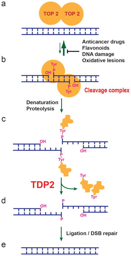
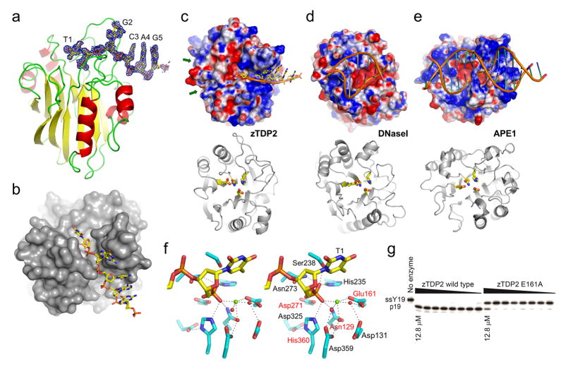
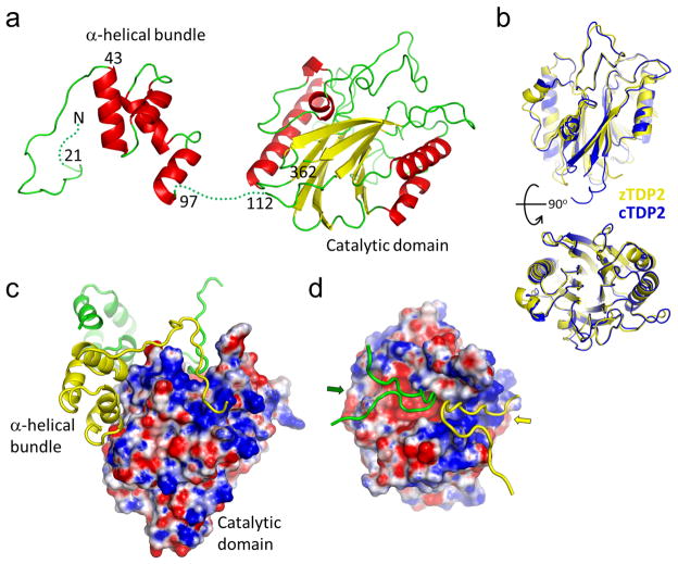
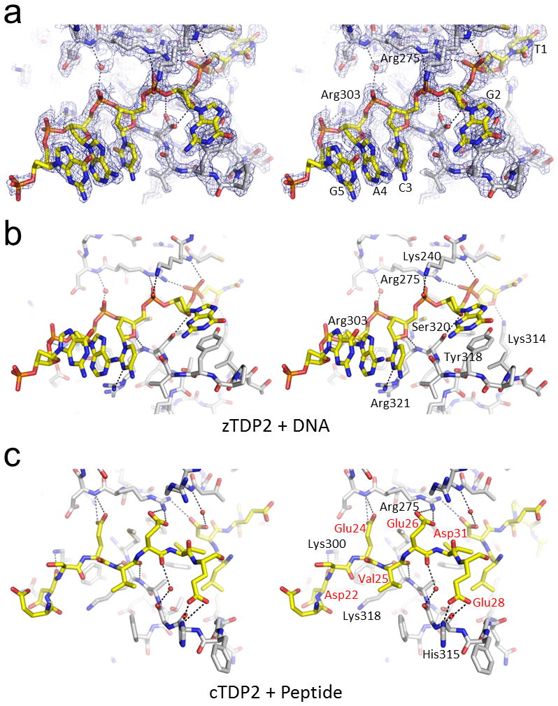
The DNA-repair enzyme Tdp2 resolves 5'-phosphotyrosyl DNA adducts and mediates resistance to anticancer drugs that target covalent topoisomerase-DNA complexes. Tdp2 also participates in key signaling pathways during development and tumorigenesis and cleaves a protein-RNA linkage during picornavirus replication. The crystal structure of zebrafish Tdp2 bound to DNA reveals a deep, narrow basic groove that selectively accommodates the 5' end of single-stranded DNA in a stretched conformation. The crystal structure of the full-length Caenorhabditis elegans Tdp2 shows that this groove can also accommodate an acidic peptide stretch in vitro, with glutamate and aspartate side chains occupying the DNA backbone phosphate-binding sites. This extensive molecular mimicry suggests a potential mechanism for autoregulation and interaction of Tdp2 with phosphorylated proteins in signaling. Our study provides a framework to interrogate functions of Tdp2 and develop inhibitors for chemotherapeutic and antiviral applications.


Conflict of interest statement

Competing financial interests:

The authors declare no competing financial interests.

Figures

Figure 1
Figure 1. Repair of topoisomerase II cleavage complexes by TDP2
(a, b) Topoisomerase II (TOP2) functions as a homodimer, where each monomer cleaves one strand of a double-stranded DNA by forming a covalent 5′-phosphotyrosyl bond. The resulting cleavage complex (b) allows the passage of another duplex DNA through the break, thereby enabling DNA relaxation, catenation-decatenation, and knotting-unknotting. Under normal conditions, religation of the cleaved DNA strands is highly efficient and most of TOP2 is non-covalently bound to DNA as in (a). In the presence of anticancer drugs such as etoposide, mitoxantrone, doxorubicin, and daunorubicin, or food flavonoids or DNA damage or oxidative lesions, the cleavage complex accumulates and needs to be removed for DNA repair. (c) Prior to TDP2 activity, the cleavage complex needs to be denatured or proteolyzed to expose the DNA-5′-phosphotyrosyl bond. (d) TDP2 releases the TOP2 polypeptide and leaves the 5′-phosphate end. (e) The DNA break may be repaired by direct ligation after annealing of the two ends with the 4-base pair stagger, or through double-strand break (DSB) repair mechanisms.
Figure 2
Figure 2. Structure of zTDP2, the unique mode of DNA-binding, and the active site architecture
(a) The catalytic domain of zTDP2 (ribbons) and the bound DNA (sticks), with simulated annealing composite omit 2Fo−Fc electron density contoured at 1.0σ shown for 1.8Å from the DNA atoms. (b) The molecular surface of zTDP2 with DNA bound in a narrow groove leading to the active site. (c–e) Top, surface electrostatic potential (red:−5kT e−1 to blue:+5kT e−1) of zTDP2, DNaseI, and APE1 (ref. 23) with bound DNA, in the same orientation as in the bottom panels. Bottom, backbone fold of each protein in ribbons, with sidechains for the conserved tetrad of catalytic residues (Asn, Glu, Asp, His) in sticks. The green arrows in (c) indicate the shallow grooves opposite the DNA-binding groove across the active site of zTDP2 potentially involved in binding topoisomerase-derived substrate peptides. (f) A wall-eye stereo pair showing the 5′-terminus of the single-stranded DNA and surrounding protein residues in the active site. The 4 sidechains shown in (c) are highlighted by red labels. The green sphere represents a divalent metal ion, whereas the red spheres represent water molecules. Hydrogen bonds or metal coordination interactions are denoted by dotted lines. (g) In vitro 5′-phosphotyrosine bond hydrolysis activity of the wild-type and E161A mutant zTDP2. ssY19 and p19 represent the 5′-phosphotyrosyl-oligonucleotide substrate and 5′-phospho-oligonucleotide product, respectively.
Figure 3
Figure 3. The full-length TDP2 has a modular architecture
(a) Full-length cTDP2 molecule in the crystal, with residue numbers indicated. The extreme N-terminal 20 residues as well as the linker between the α-helical bundle and the catalytic domain are disordered (dotted lines). (b) Structural comparison between the catalytic domains of zTDP2 (yellow) and cTDP (blue). Two different views (90° rotated) are shown. (c) Molecular surface of the cTDP2 catalytic domain colored according to the electrostatic potential, with the N-terminal regions of two other molecules (yellow and green) shown in ribbons. (d) Top view, similar to that of the zTDP2-DNA complex in the top panel in Fig. 2c. The basic DNA-binding groove is occupied by the N-terminal residues of one molecule (yellow), while the active site pocket and the adjacent groove are blocked by those from another molecule (green). The surface electrostatic potential is color-scaled as in Fig. 2.
Figure 4
Figure 4. Similarities between the DNA and peptide binding modes of TDP2
(a) Single-stranded DNA bound in the DNA-binding groove of zTDP2. The simulated annealing composite omit 2Fo−Fc electron density (1.0σ) is shown for 1.8Å from DNA or protein atoms or a water molecule involved in the protein-DNA interaction. (b) zTDP2-DNA interactions as in (a), from a slightly rotated view. Only one of the two conformers for the Ser320 sidechain with higher occupancy is shown for clarity. (c) The N-terminal acidic residues of cTDP2 bound in the DNA-binding groove. Wall-eye stereo pairs are shown for (a–c). Hydrogen bonding and salt bridge interactions are indicated by the black dotted lines.
Figure 5
Figure 5. cTDP2 dimerization by domain swapping
(a) Selected molecules within the crystal lattice are shown in different colors to illustrate the intermolecular interactions. The asymmetric unit contains one cTDP2 molecule in the full-length cTDP2 crystal, and thus all molecules are crystallographically equivalent. (b) The α-helical bundle (yellow tube) interacting with the relatively hydrophobic (white) surface of the catalytic domain. Molecular surface of the catalytic domain is colored according to the electrostatic potential scaled as in Fig. 2. (c) Size-exclusion chromatography profiles for cTDP2 and its deletion mutants. Positions of the molecular weight standards are indicated by arrows. (d) SAXS analysis shows that the N-terminal residues mediate dimer formation in full-length cTDP2 in solution. SAXS curves for the full-length cTDP2 and cTDP2ΔN107, with linear Guinier plots (inset) showing the lack of aggregation in the sample. (e) GASBOR ab inito shape predictions are consistent with a monomeric form for cTDP2ΔN107 and dimeric form for full-length cTDP2. (f) In vitro 5′-phosphotyrosine bond hydrolysis activity of the full-length and truncated cTDP2. ssY19 and p19 represent the 5′-phosphotyrosyl-oligonucleotide substrate and 5′-phospho-oligonucleotide product, respectively.

Comment in

References

    1. Pommier Y, Leo E, Zhang H, Marchand C. DNA topoisomerases and their poisoning by anticancer and antibacterial drugs. Chem Biol. 2010;17:421–433. - PMC - PubMed
    1. Wang JC. Cellular roles of DNA topoisomerases: a molecular perspective. Nat Rev Mol Cell Biol. 2002;3:430–440. - PubMed
    1. Champoux JJ. DNA topoisomerases: structure, function, and mechanism. Annu Rev Biochem. 2001;70:369–413. - PubMed
    1. Corbett KD, Berger JM. Structure, molecular mechanisms, and evolutionary relationships in DNA topoisomerases. Annu Rev Biophys Biomol Struct. 2004;33:95–118. - PubMed
    1. Haffner MC, et al. Androgen-induced TOP2B-mediated double-strand breaks and prostate cancer gene rearrangements. Nat Genet. 2010;42:668–675. - PMC - PubMed

Publication types