. 2012 Dec;13(12):1178-86.
doi: 10.1038/ni.2457. Epub 2012 Oct 28.

Immunodeficiency, autoinflammation and amylopectinosis in humans with inherited HOIL-1 and LUBAC deficiency

Affiliations

Immunodeficiency, autoinflammation and amylopectinosis in humans with inherited HOIL-1 and LUBAC deficiency

Bertrand Boisson et al. Nat Immunol. 2012 Dec.

Abstract

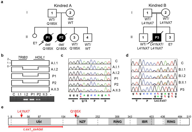
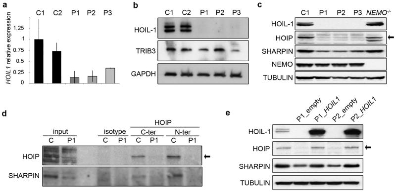
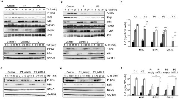
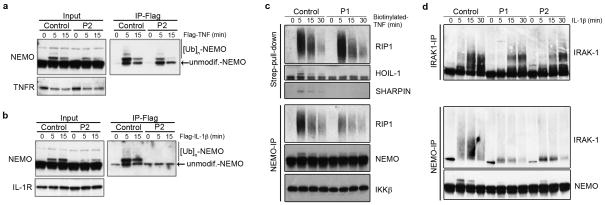
We report the clinical description and molecular dissection of a new fatal human inherited disorder characterized by chronic autoinflammation, invasive bacterial infections and muscular amylopectinosis. Patients from two kindreds carried biallelic loss-of-expression and loss-of-function mutations in HOIL1 (RBCK1), a component of the linear ubiquitination chain assembly complex (LUBAC). These mutations resulted in impairment of LUBAC stability. NF-κB activation in response to interleukin 1β (IL-1β) was compromised in the patients' fibroblasts. By contrast, the patients' mononuclear leukocytes, particularly monocytes, were hyper-responsive to IL-1β. The consequences of human HOIL-1 and LUBAC deficiencies for IL-1β responses thus differed between cell types, consistent with the unique association of autoinflammation and immunodeficiency in these patients. These data suggest that LUBAC regulates NF-κB-dependent IL-1β responses differently in different cell types.


Conflict of interest statement

Competing interest statement: The authors have no competing financial interests to declare.

Figures

Figure 1
Figure 1. Two kindreds with autosomal recessive HOIL1 deficiency
a) Pedigree of kindred A, showing the segregation of the 31.799 kb deletion of chromosome 20 (del: TRIB3:g.-1272_HOIL1:g.9780del) encompassing TRIB3 and HOIL1, and the nonsense c.553 C>T, p.Q185X mutant allele in HOIL1. Pedigree of kindred B, showing the segregation of the HOIL1 deletion c.121_122delCT, p.L41fsX7. The arrow indicates the index case. b) Schematic representation of the deletion encompassing the two genes, with the loss of one copy of the allele from individuals A.I.2, P1 and P2. In the lower panel, a PCR-based approach involving amplification of a 1.235 kb fragment with genomic TRIB3 and HOIL1 primers reveals the deletion. c–d) HOIL1 DNA sequence electropherograms, for a control and the patients c) from kindred A, for the region corresponding to the nonsense mutation and d) from kindred B, for the region corresponding to the deletion. e) Schematic diagram of the HOIL-1 protein. Ubiquitin-like (Ubl), novel zinc-finger (NZF), ring (RING) and in-between RING (IBR) domains are shaded in gray. Arrows indicate the nonsense and deletion mutations and the double arrow indicates the deletion of the first four exons in HOIL1. Exon boundaries are indicated by vertical dashed bars and amino-acid positions are numbered.
Figure 2
Figure 2. HOIL-1 complete deficiency
a) Relative expression of HOIL1 in SV40-immortalized fibroblasts from controls (C1 and C2) and patients (P1, P2 and P3). b) HOIL-1 and TRIB3 immunoblots of total cell extracts from SV40-immortalized fibroblasts derived from controls and patients. c) Low levels of HOIP and SHARPIN in the absence of HOIL-1 in SV40-immortalized fibroblasts. d) No HOIP-SHARPIN co-immunoprecipitates can be detected in HOIL-1-deficient fibroblasts. Immunoprecipitation experiments were performed with either control mouse IgGs (isotype) or mouse antibodies against the N-terminus or C-terminus of HOIP in control or P1 patient-derived fibroblasts. HOIP and co-immunoprecipitated SHARPIN were detected by immunoblotting. e) Re-expression of HOIL-1 in fibroblasts from patients restores HOIP and SHARPIN protein levels. A retrovirus-based strategy was used for stable re-expression of the wt HOIL1 allele in fibroblasts from P1 and P2. Fibroblasts infected with empty viruses were used as controls. Data are representative of three experiments (a), four experiments (b), three experiments (c), three experiments (d) or four experiments (e).
Figure 3
Figure 3. HOIL-1 is required for full TNF-α and IL-1β-induced activation of NF-κB in HOIL-1-deficient fibroblasts
a) Impaired NF-κB activation in response to TNF. Time-course of TNF-stimulated SV40-immortalized fibroblasts, showing impaired IKK kinase phosphorylation, lower levels of NEMO ubiquitination (indicated by an asterisk) and slower IκBα degradation in patients than in controls. JNK phosphorylation in response to TNF is identical in control and patient cells. b) Impaired NF-κB activation in response to IL-1β. Time-course of IL-1β-stimulated SV40-immortalized fibroblasts showing impaired IKK kinase phosphorylation (upper panel), lower levels of NEMO ubiquitination (indicated by an asterisk) and slower IκBα degradation in patients than in the controls. JNK phosphorylation in response to IL-1β is identical in control and patient cells. c) Low levels of IL-6 production in response to TNF and IL-1β in SV40-immortalized fibroblasts from HOIL-1-deficient patients (P1, P2, P3). All numerical data are means ± SEM. * p<0.05, ** p<0.01, *** p<0.001, ns, not significant. d–e) Re-expression of HOIL-1 restores the TNF- and IL-1β-induced activation of NF-κB. The kinetics of TNF and IL-1β stimulation in the indicated cell lines was followed by determining the levels of phospho-IKKs, IκB α and NEMO by immunoblotting. GAPDH was used as a control. f) HOIL-1 complementation restores TNF-and IL-1β-induced IL-6 production in fibroblasts from P1 and P2. IL-6 levels in the supernatants of mock-, TNF- and IL-1β-treated cell lines were assayed by ELISA. All numerical data are means ± SEM. (** p<0.01). Data are representative of three experiments (a,b), three experiments (c), two experiments (d,e) or three experiments (f).
Figure 4
Figure 4. Impaired recruitment of NEMO to cytokine receptors in the patients’ fibroblasts
a) HOIL-1 deficiency impairs the recruitment of NEMO to the TNF-RSC. Flag-tagged TNF was used to activate and isolate the TNF-RSC. Cells were lysed, TNF-RSC was purified on Flag-affinity resin and complex-associated NEMO was analyzed by immunoblotting. b) HOIL-1 deficiency abolishes NEMO recruitment to the IL-1-RSC. The same strategy as in a) was used, except that Flag-tagged IL-1β was used to stimulate the cells and to isolate the IL-1-RSC. The abundance of TNFR (a) and IL-1R (b), assessed by immunoblotting, was similar in control and patient-derived fibroblasts. c) HOIL-1 deficiency impairs the interaction between NEMO and polyubiquitinated RIP1 in response to TNF. NEMO (NEMO-IP) was immunoprecipitated from lysates of control and patient fibroblasts treated with biotinylated TNF and analyzed by immunoblotting for NEMO, RIP1, and IKKβ. The total amount of the ubiquitinated forms of RIP1 associated with the TNF-RSC was evaluated by streptavidin pulldown followed by immunoblotting for RIP1. d) HOIL-1 deficiency impairs the interaction between NEMO and polyubiquitinated IRAK-1 in response to IL-1β. NEMO and IRAK-1 were immunoprecipitated from lysates of IL-1β-treated fibroblasts and subjected to western blotting for NEMO or IRAK-1 as indicated. These data are representative of three experiments.
Figure 5
Figure 5. Transcriptome analysis of TNF or IL-1β stimulation of primary fibroblasts
Primary fibroblasts from four healthy donors, P1, P2, P3, MyD88- and NEMO-deficient cells were stimulated with TNF or IL-1β for 2 and 6 hours. Stimulated samples were normalized with respect to the corresponding non stimulated reference sample. The heat maps represent hierarchically clustered transcripts displaying up- or downregulation by a factor of at least two in stimulated healthy control samples (averaged).
Figure 6
Figure 6. Whole blood analysis reveals a new hyperinflammatory disorder in HOIL-1-deficient patients
a) Transcriptional profiles of whole blood from patients with hyperinflammatory conditions (CINCA, MWS, MVK) and HOIL-1-deficient patients. Comparison of the transcriptional profile of P1 with those of 41 healthy age-matched children (not shown) and other patients with CAPS (2 CINCA, 5 MWS and 2 MVK patients). P1 presented a distinct pattern of gene expression in the blood, with 2,900 transcripts up- or downregulated by a factor of more than two with respect to healthy controls. Red, blue and yellow indicate a relative increase, decrease and no change in expression levels respectively. Samples are ordered by donor: HOIL-1-deficient patient (P1, lane 1), CINCA patients (lanes 2–3), MWS patients (lanes 4–8) and MVK-deficient patients (lanes 9–10). b) Quantification of various cytokines in plasma samples. The amounts of IL-1RA (an IL-1 antagonist), TNF, IL-6 and CRP were determined in plasma samples from healthy donors, P1 and P2 taken at various ages. Individuals with CRP concentrations above 3 mg/ml were considered to have an inflammatory condition. c) IL-6 secretion by whole-blood cells from 30 healthy donors and HOIL-1-deficient patients (P1, P2), treated with IL-1β for 48 hours. All numerical data are means ± SEM, *** p<0.001 d) IL-10 secretion by whole-blood cells from healthy donors and HOIL-1-deficient patients (P1, P2) and the heterozygous parents (I.1, I.2), activated by incubation with TNF or PMA+ionomycin for 48 hours. nd: not detectable
Figure 7
Figure 7. HOIL-1-deficient monocytes display hyperproduction of IL-6 upon IL-1β stimulation
a) Transcriptional profile of leukocytes from P1, P2 and P3 after 2 and 6 hours of stimulation by TNF or IL-1β. The median gene expression in HOIL-1- or MyD88-deficient patients (y-axis) was plotted against the median gene expression sorted by ascending order of fold induction in the healthy controls (two of whom were age-matched). Each dot corresponds to one probe. b) Levels of IL6, IL8, MIP1A, MIP1B and IL1B mRNA in 3 healthy controls, 3 HOIL-1- (P1, P2 and P3) and 2 MyD88-deficient patients, extracted from the microarray data. Each dot corresponds to one probe in one individual. The number of probes per gene is 1 for IL6 and IL1B, 2 for MIP1B and IL8 and 4 for MIP1A. The non parametric Mann-Whitney test was used for statistical analysis (* p<0.1, ** p<0.05, *** p<0.005). c) Cytokine production in CD14+ cell subsets determined by intracellular staining of stimulated PBMCs. PBMCs from P2, 7 healthy controls and 1 IRAK-4-deficient patient were stimulated with TNF, IL-1β or LPS. The secretory pathways were concomitantly blocked with monensin and brefeldin A. Twelve hours after stimulation, the cells were immunolabeled with antibodies against CD14 and then permeabilized to assess the production of IL-6, MIP-1α and IL-8. Cells were analyzed by flow cytometry. (n=1).

Comment in

References

    1. Masters SL, Simon A, Aksentijevich I, Kastner DL. Horror autoinflammaticus: the molecular pathophysiology of autoinflammatory disease (*) Annu Rev Immunol. 2009;27:621–668. - PMC - PubMed
    1. Aksentijevich I, et al. An autoinflammatory disease with deficiency of the interleukin-1-receptor antagonist. N Engl J Med. 2009;360:2426–2437. - PMC - PubMed
    1. McDermott MF, et al. Germline mutations in the extracellular domains of the 55 kDa TNF receptor, TNFR1, define a family of dominantly inherited autoinflammatory syndromes. Cell. 1999;97:133–144. - PubMed
    1. Puel A, Picard C, Ku CL, Smahi A, Casanova JL. Inherited disorders of NF-kappaB-mediated immunity in man. Curr Opin Immunol. 2004;16:34–41. - PubMed
    1. Doffinger R, et al. X-linked anhidrotic ectodermal dysplasia with immunodeficiency is caused by impaired NF-kappaB signaling. Nat Genet. 2001;27:277–285. - PubMed

Publication types

MeSH terms

Associated data