

Genomic aberrations in normal tissue adjacent to HER2-amplified breast cancers: field cancerization or contaminating tumor cells?
- PMID: 23104223
- PMCID: PMC3511697
- DOI: 10.1007/s10549-012-2290-3
Genomic aberrations in normal tissue adjacent to HER2-amplified breast cancers: field cancerization or contaminating tumor cells?
Abstract
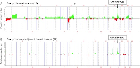
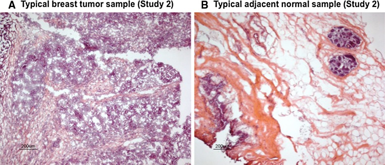
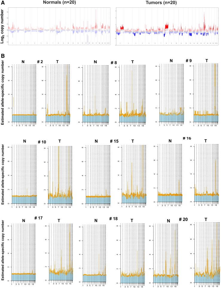
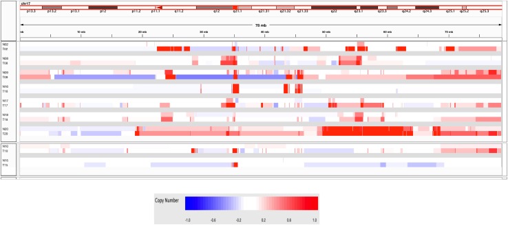
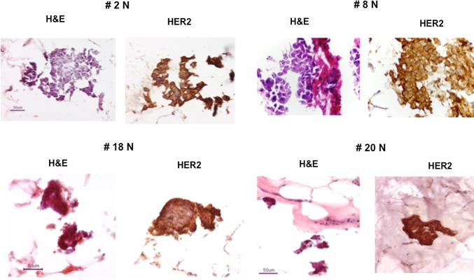
Field cancerization effects as well as isolated tumor cell foci extending well beyond the invasive tumor margin have been described previously to account for local recurrence rates following breast conserving surgery despite adequate surgical margins and breast radiotherapy. To look for evidence of possible tumor cell contamination or field cancerization by genetic effects, a pilot study (Study 1: 12 sample pairs) followed by a verification study (Study 2: 20 sample pairs) were performed on DNA extracted from HER2-positive breast tumors and matching normal adjacent mammary tissue samples excised 1-3 cm beyond the invasive tumor margin. High-resolution molecular inversion probe (MIP) arrays were used to compare genomic copy number variations, including increased HER2 gene copies, between the paired samples; as well, a detailed histologic and immunohistochemical (IHC) re-evaluation of all Study 2 samples was performed blinded to the genomic results to characterize the adjacent normal tissue composition bracketing the DNA-extracted samples. Overall, 14/32 (44 %) sample pairs from both studies produced genome-wide evidence of genetic aberrations including HER2 copy number gains within the adjacent normal tissue samples. The observed single-parental origin of monoallelic HER2 amplicon haplotypes shared by informative tumor-normal pairs, as well as commonly gained loci elsewhere on 17q, suggested the presence of contaminating tumor cells in the genomically aberrant normal samples. Histologic and IHC analyses identified occult 25-200 μm tumor cell clusters overexpressing HER2 scattered in more than half, but not all, of the genomically aberrant normal samples re-evaluated, but in none of the genomically normal samples. These genomic and microscopic findings support the conclusion that tumor cell contamination rather than genetic field cancerization represents the likeliest cause of local clinical recurrence rates following breast conserving surgery, and mandate caution in assuming the genomic normalcy of histologically benign appearing peritumor breast tissue.
Figures
References
-
- Carlson RW, Allred DC, Anderson BO, et al. Invasive breast cancer. J Natl Compr Cancer Netw. 2011;9:136–222. - PubMed
-
- Shar JP, Rosen PP, Robbins GF. Pitfalls of local excision in the treatment of carcinoma of the breast. Surg Gynecol Obstet. 1973;136:721–725. - PubMed
-
- Morgenstern L, Friedmann NB. Breast cancer: the case against tylectomy. The factor of multicentricity. Prog Clin Cancer. 1978;76:113–122. - PubMed
-
- Lagios MD, Westdahl PR, Rose MR. The concept and implications of multicentricity in breast carcinoma. Pathol Annu. 1981;16:83–102. - PubMed
Publication types
MeSH terms
Substances
Grants and funding
LinkOut - more resources
Full Text Sources
Medical
Research Materials
Miscellaneous
