. 2013 Jan;79(1):303-13.
doi: 10.1128/AEM.02761-12. Epub 2012 Oct 26.

Phylogenetic, metabolic, and taxonomic diversities shape mediterranean fruit fly microbiotas during ontogeny

Affiliations

Phylogenetic, metabolic, and taxonomic diversities shape mediterranean fruit fly microbiotas during ontogeny

Yael Aharon et al. Appl Environ Microbiol. 2013 Jan.

Abstract

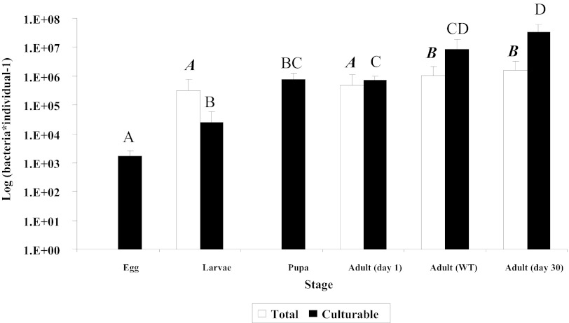
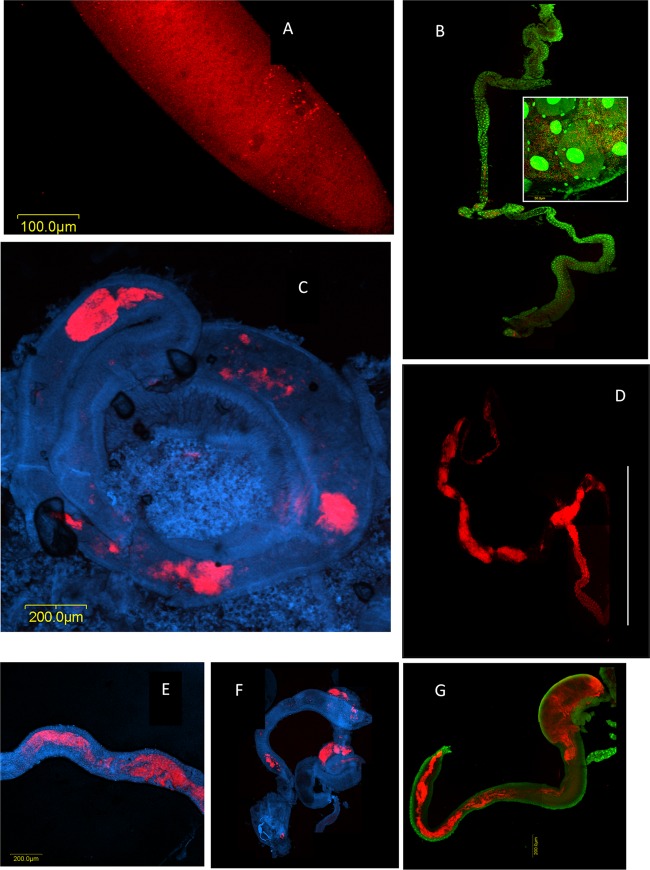
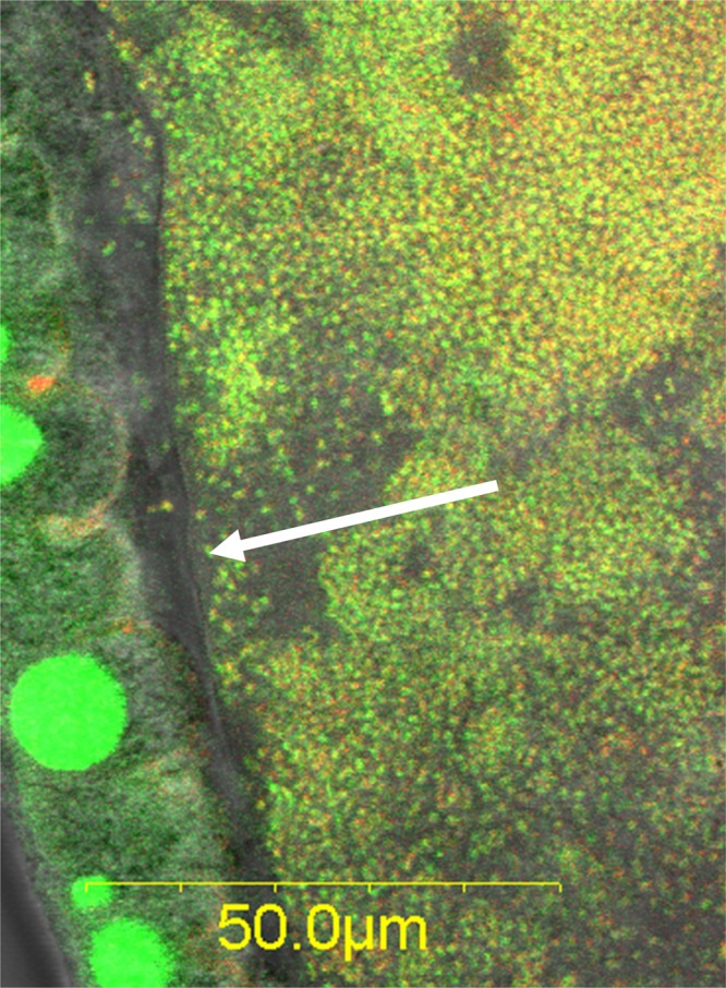
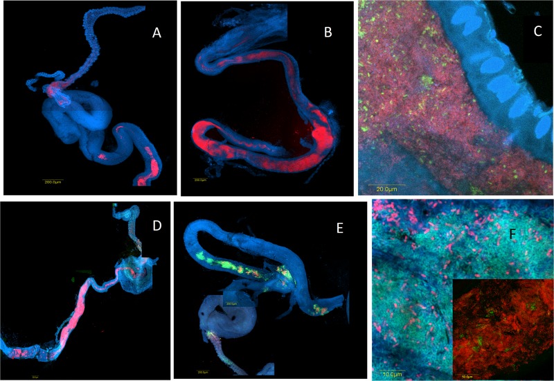
The Mediterranean fruit fly (medfly) (Ceratitis capitata) lays eggs in fruits, where larvae subsequently develop, causing large-scale agricultural damage. Within its digestive tract, the fly supports an extended bacterial community that is composed of multiple strains of a variety of enterobacterial species. Most of these bacteria appear to be functionally redundant, with most strains sustaining diazotrophy and/or pectinolysis. At least some of these bacteria were shown to be vertically inherited, but colonization, structural, and metabolic aspects of the community's dynamics have not been investigated. We used fluorescent in situ hybridization, metabolic profiling, plate cultures, and pyrosequencing to show that an initial, egg-borne, diverse community expands throughout the fly's life cycle. While keeping "core" diazotrophic and pectinolytic functions, it also harbors diverse and fluctuating populations that express varied metabolic capabilities. We suggest that the metabolic and compositional plasticity of the fly's microbiota provides potential adaptive advantages to the medfly host and that its acquisition and dynamics are affected by mixed processes that include stochastic effects, host behavior, and molecular barriers.


Figures

Fig 1
Fig 1
Culturable and total bacteria per individual medfly during consecutive developmental stages. Black and white columns represent culturable bacteria, as enumerated by dilution plating, and total bacteria, as counted by microscopy, respectively. Midgut bacteria in adults and larvae were counted. Developmental stages not connected by the same letter are significantly different. Boldface italic type refers to white columns, and lightface type refers to black columns. Error bars represent standard deviations.
Fig 2
Fig 2
Bacterial colonization of the medfly during ontogeny, tracked by fluorescent in situ hybridization and confocal microscopy using a Bacteria-directed Cy3-EUB338 oligonucleotide probe applied on laboratory-raised flies and DAPI or Sybr green nucleic acid stain. (A) Egg. Microcolonies on the surface of a medfly egg appear as red dots on the red fluorescent background of the egg chorion. (B) Larval gut. Bacterial growth appears as discontinuous patches along the larval gut. Red, bacterial masses; green, fly gut cell nuclei. (Inset) Within these patches, the bacterial density is high. Red, bacterial cells/microcolonies; green, bacterial cells and fly gut cell nuclei. (C) Adult medfly midgut 1 day after eclosion. Red, bacterial masses. (D) Midgut and hindgut of a laboratory-reared female adult medfly 30 days after eclosion. Continuous bacterial colonization is seen in the midgut as well as in the hindgut. Red, bacterial masses. The line encompasses the hindgut. (E to G) Midguts of field-captured wild specimens showing various extents of gut colonization. (E) Female; (F and G) males. Red, bacterial masses; blue or green, gut cells.
Fig 3
Fig 3
The microbiota is contained in the gut lumen by the peritrophic membrane (arrow), as seen in a wild male. Green, Sybr green nucleic acid stain; red, Cy3-EUB338 Bacteria-specific probe. Bacteria stained with both Sybr green and Cy3 appear yellow. Large green spots are epithelial cell nuclei.
Fig 4
Fig 4
Colonization of laboratory-reared flies at 13 days (A and C) and 30 days (B and D) following eclosion and of wild-caught flies (E and F) with bacterial populations hybridizing with oligonucleotide probes. (A and B) Klebsiella (Cy3-Kox615) (red); (C) Enterobacter (FAM6-Ent615) (green) and Bacteria (Cy3-EUB3380 (red); (D to F) Enterobacter (FAM6-Ent615) (green) and Klebsiella (Cy3-Kox615) (red). Blue indicates staining by DAPI (nucleic acid stain).

References

    1. Weinert LA, Tinsley MC, Temperley M, Jiggins FM. 2007. Are we underestimating the diversity and incidence of insect bacterial symbionts? A case study in ladybird beetles. Biol. Lett. 3:678–681 - PMC - PubMed
    1. Baumann P, Moran NA, Baumann L. 2006. Bacteriocyte-associated endosymbionts of insects, p 403–438 In Dworkin M, Rosenberg E, Schleifer K-H, Stackebrandt E. (ed), The prokaryotes. Springer, New York, NY
    1. Behar A, Jurkevitch E, Yuval B. 2008. Bringing back the fruit into fruit fly-bacteria interactions. Mol. Ecol. 17:1375–1386 - PubMed
    1. Ben Yosef M, Jurkevitch E, Yuval B. 2008. Effect of bacteria on nutritional status and reproductive success of the Mediterranean fruit fly Ceratitis capitata. Physiol. Entomol. 33:145–154
    1. Kaiwa N, Hosokawa T, Kikuchi Y, Nikoh N, Meng XY, Kimura N, Ito M, Fukatsu T. 2010. Primary gut symbiont and secondary, Sodalis-allied symbiont of the scutellerid stinkbug Cantao ocellatus. Appl. Environ. Microbiol. 76:3486–3494 - PMC - PubMed

Publication types

LinkOut - more resources