

Do cartilage repair procedures prevent degenerative meniscus changes?: longitudinal t1ρ and morphological evaluation with 3.0-T MRI
- PMID: 23104606
- PMCID: PMC4074395
- DOI: 10.1177/0363546512461594
Do cartilage repair procedures prevent degenerative meniscus changes?: longitudinal t1ρ and morphological evaluation with 3.0-T MRI
Abstract
Background: Cartilage repair (CR) procedures are widely accepted for treatment of isolated cartilage defects in the knee joint. However, it is not well known whether these procedures prevent degenerative joint disease.
Hypothesis: Cartilage repair procedures prevent accelerated qualitative and quantitative progression of meniscus degeneration in individuals with focal cartilage defects.
Study design: Cohort study; Level of evidence, 2.
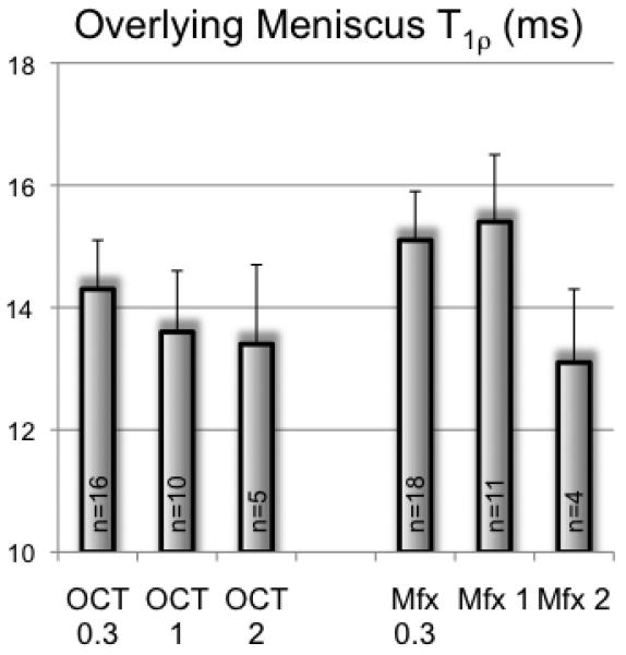
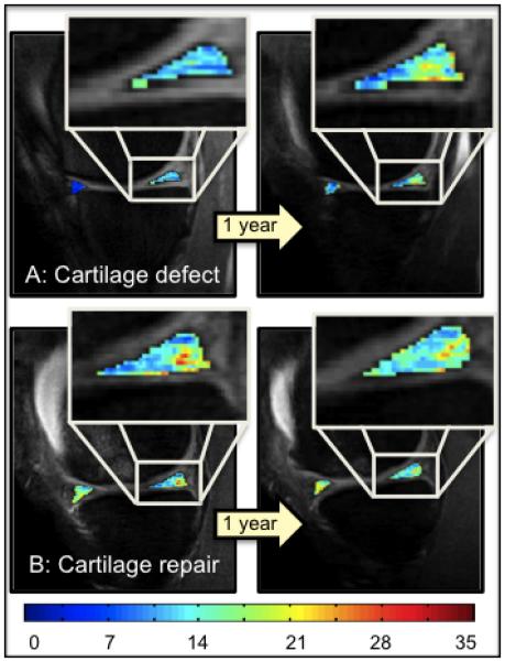
Methods: Ninety-four subjects were studied. Cartilage repair procedures were performed on 34 patients (osteochondral transplantation, n = 16; microfracture, n = 18); 34 controls were matched. An additional 13 patients received CR and anterior cruciate ligament (ACL) reconstruction (CR&ACL), and 13 patients received only ACL reconstruction. Magnetic resonance imaging at 3.0-tesla with T(1ρ) mapping and sagittal fat-saturated intermediate-weighted fast spin echo (FSE) sequences was performed to quantitatively and qualitatively analyze menisci (Whole-Organ Magnetic Resonance Imaging Score [WORMS] assessment). Patients in the CR and CR&ACL groups were examined 4 months (n = 34; n = 13), 1 year (n = 21; n = 8), and 2 years (n = 9; n = 5) after CR. Control subjects were scanned at baseline and after 1 and 2 years, ACL patients after 1 and 2 years.
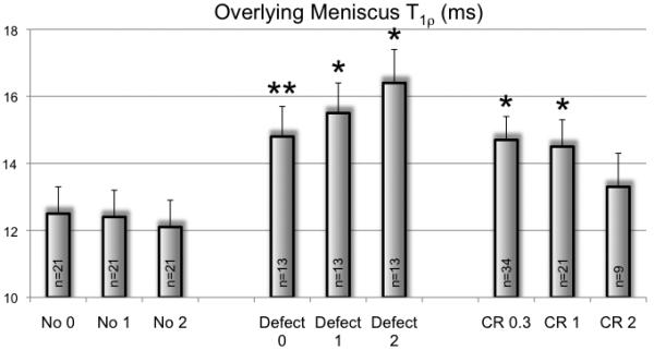
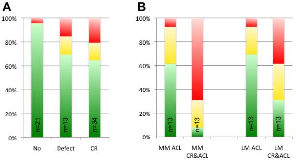
Results: At baseline, global meniscus T(1ρ) values (mean ± SEM) were higher in individuals with CR (14.2 ± 0.5 ms; P = .004) and in individuals with CR&ACL (17.1 ± 0.9 ms; P < .001) when compared with controls (12.8 ± 0.6 ms). After 2 years, there was a statistical difference between T(1ρ) at the overlying meniscus above cartilage defects (16.4 ± 1.0 ms) and T(1ρ) of the subgroup of control knees without cartilage defects (12.1 ± 0.8 ms; P < .001) and a statistical trend to the CR group (13.3 ± 1.0 ms; P = .09). At baseline, 35% of subjects with CR showed morphological meniscus tears at the overlying meniscus; 10% of CR subjects showed an increase in the WORMS meniscus score within the first year, and none progressed in the second year. Control subjects with (without) cartilage defects showed meniscus tears in 30% (5%) at baseline; 38% (19%) increased within the first year, and 15% (10%) within the second year.
Conclusion: This study demonstrated more severe meniscus degeneration after CR surgery compared with controls. However, progression of T(1ρ) values was not observed from 1 to 2 years after surgery. These results suggest that CR may prevent degenerative meniscus changes.
Figures

References
-
- Akella SV, Regatte RR, Gougoutas AJ, et al. Proteoglycan-induced changes in T1rhorelaxation of articular cartilage at 4T. Magn Reson Med. 2001;46(3):419–423. - PubMed
-
- Bedi A, Feeley BT, Williams RJ., 3rd Management of articular cartilage defects of the knee. J Bone Joint Surg Am. 2010;92(4):994–1009. - PubMed
-
- Bellabarba C, Bush-Joseph CA, Bach BR., Jr. Patterns of meniscal injury in the anterior cruciate-deficient knee: a review of the literature. Am J Orthop (Belle Mead NJ) 1997;26(1):18–23. - PubMed
-
- Benthien JP, Schwaninger M, Behrens P. We do not have evidence based methods for the treatment of cartilage defects in the knee. Knee Surg Sports Traumatol Arthrosc. 2011;19(4):543–552. - PubMed
