. 2013 Feb;27(2):557-67.
doi: 10.1096/fj.12-218156. Epub 2012 Oct 26.

The prostamide-related glaucoma therapy, bimatoprost, offers a novel approach for treating scalp alopecias

Affiliations

The prostamide-related glaucoma therapy, bimatoprost, offers a novel approach for treating scalp alopecias

Karzan G Khidhir et al. FASEB J. 2013 Feb.

Abstract

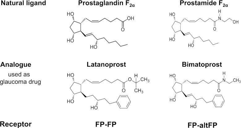
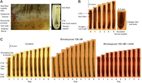
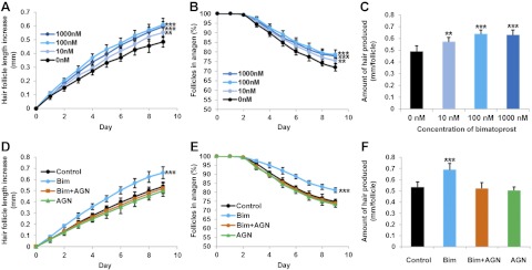
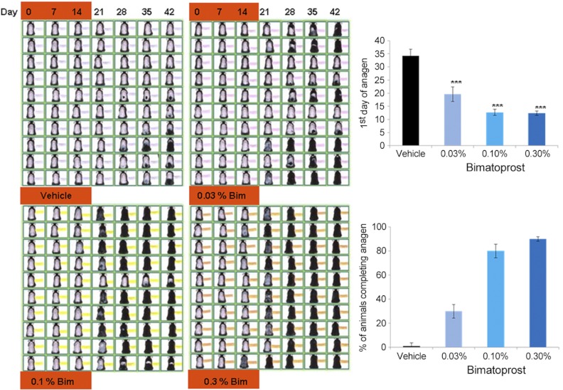
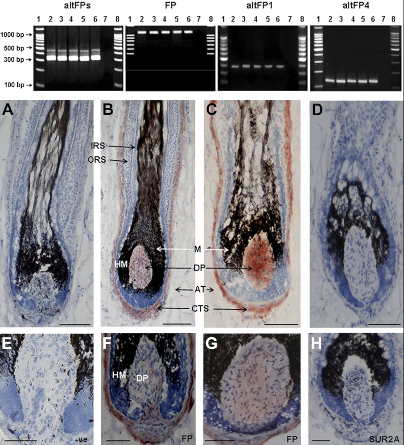
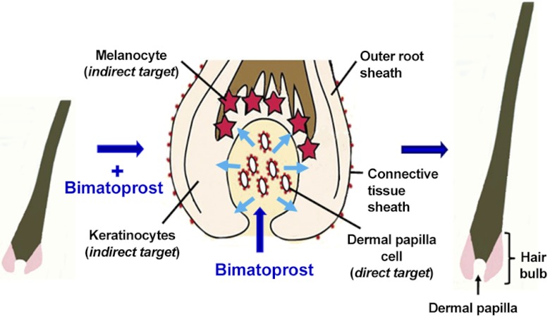
Balding causes widespread psychological distress but is poorly controlled. The commonest treatment, minoxidil, was originally an antihypertensive drug that promoted unwanted hair. We hypothesized that another serendipitous discovery, increased eyelash growth side-effects of prostamide F(2α)-related eyedrops for glaucoma, may be relevant for scalp alopecias. Eyelash hairs and follicles are highly specialized and remain unaffected by androgens that inhibit scalp follicles and stimulate many others. Therefore, we investigated whether non-eyelash follicles could respond to bimatoprost, a prostamide F(2α) analog recently licensed for eyelash hypotrichosis. Bimatoprost, at pharmacologically selective concentrations, increased hair synthesis in scalp follicle organ culture and advanced mouse pelage hair regrowth in vivo compared to vehicle alone. A prostamide receptor antagonist blocked isolated follicle growth, confirming a direct, receptor-mediated mechanism within follicles; RT-PCR analysis identified 3 relevant receptor genes in scalp follicles in vivo. Receptors were located in the key follicle regulator, the dermal papilla, by analyzing individual follicular structures and immunohistochemistry. Thus, bimatoprost stimulates human scalp follicles in culture and rodent pelage follicles in vivo, mirroring eyelash behavior, and scalp follicles contain bimatoprost-sensitive prostamide receptors in vivo. This highlights a new follicular signaling system and confirms that bimatoprost offers a novel, low-risk therapeutic approach for scalp alopecias.


Figures

Figure 1.
Figure 1.
Comparison of PGF and prostamide F, their analogs, and receptors. The prostamides are electrochemically neutral biological lipids related to prostaglandins, but with a terminal ethanolamide group. PGF-related analogs, e.g., latanoprost, and the prostamide F-related analog, bimatoprost, are used for glaucoma. Alternative splicing variants of FP appear to heterodimerize with FP to create a bimatoprost-sensitive prostamide receptor (30).
Figure 2.
Figure 2.
Sequential photomicrographs of human scalp hair follicles growing in organ culture. A) Isolation of hair follicles from scalp skin. Left panel: scalp skin, showing hair follicles and skin layers. Right panel: isolated hair follicle, demonstrating the hair fiber, hair matrix (pigmented in the upper hair bulb and unpigmented below), dermal papilla, and CTS. B, C) Sequential photomicrographs taken every 24 h for 9 d of individual scalp follicles in organ culture under various conditions showing growth of hair fiber, but not the CTS. B) Some follicles showed catagen-like changes in hair bulb morphology. By d 5, pigmentation had ceased, and the base of the hair fiber was retracting upward, leaving behind a ball of dermal papilla cells. C) Most follicles maintained bulb anagen morphology and increased in length over 9 d. Follicles were cultured in control medium (left), 100 nM bimatoprost (center), or 100 nM bimatoprost and AGN 211336 (AGN; 1 μM; right). Scale bars = 200 μm (A, B); 500 μm (C).
Figure 3.
Figure 3.
Bimatoprost stimulated scalp hair follicle growth in organ culture, and a prostamide F receptor antagonist blocked this stimulation. Anagen follicles were assessed daily for morphological changes and measured, while cultured with either vehicle alone (control), or bimatoprost (10, 100, and 1000 nM) with or without a specific antagonist. Values are means ± se of 5 individuals for each experiment; ≥6 follicles/person were examined for each condition. In control medium, hair follicles increased in length (A, D); the number of follicles remaining in anagen gradually declined (B, E). Bimatoprost increased scalp follicle growth rate at 10 nM (P<0.01) and 100 and 1000 nM (P<0.001) (A), number of anagen follicles at 10 nM (P<0.01) and 100 and 1000 nM (P<0.001) (B), and the total amount of hair produced at 10 nM (P<0.01) and 100 and 1000 nM (P<0.001) (C). The FP receptor antagonist AGN 211336 at 1 μM abolished the stimulatory effects of 100 nM bimatoprost on all parameters of scalp follicle growth in organ culture (P<0.01; D–F). *P < 0.05, **P < 0.01, ***P < 0.001 vs. control.
Figure 4.
Figure 4.
Topical application of bimatoprost promoted hair growth in mice in vivo. Bimatoprost applied topically to female mice for 2 wk stimulated resting (telogen) follicles to start to grow i.e., enter the next hair cycle (anagen). Pink skin visible in d 0 photographs confirms telogen status. Growing hairs are initially seen as darkening skin until black hair projects outside the skin (as seen in sequential photographs of individual animals). Bimatoprost (Bim) at 0.03, 0.10, and 0.30% significantly (P<0.001) advanced the first day at which anagen was visible, compared to the vehicle alone (top graph) and increased the percentage of animals in which all follicles on the back had grown at d 42 (bottom graph). n = 10/treatment. ***p < 0.001 vs. control.
Figure 5.
Figure 5.
Human scalp anagen hair follicles expressed the genes and protein for three prostanoid receptors. Top panel: agarose gel electrophoresis of PCR products (30 μl) of hair follicle cDNAs from isolated scalp hair follicles from each of 5 individuals showed bands corresponding to 3 prostanoid receptors, with a primer pair for native FP and all known altFPs. These 3 bands correspond to the native FP receptor (∼321 bp) and 2 larger altFPs that include the same parts of the native FP sequence plus the relevant intron sequences. Specific primers for FP (1080 bp), altFP1 (392 bp), and altFP4 (141 bp) gave appropriately sized bands. AltFP2, altFP3, and altFP5 were not detected (data not shown). Lanes 1 and 8, DNA ladder (100-1500 bp); lanes 2–6, PCR products from 5 follicle cDNAs; lane 7, negative control without cDNA. Bottom panels: immunohistochemical analysis of scalp cryosections localized FP protein in the hair bulb in the dermal papilla and CTS with two primary antibodies for human FP (B, C, F, G). No staining occurred with omission of the primary antibody (A, E) nor with the nonrelevant antibody (D, H). Normal dark pigment (melanin) is visible in the hair bulb. DP, dermal papilla; CTS, connective tissue sheath; HM, hair matrix. Red, positive staining; blue, hematoxylin counterstain. Scale bars = 150 μm (A–D); 100 μm (E–H).
Figure 6.
Figure 6.
Localization of three prostanoid receptor genes only in the scalp hair follicle dermal papilla and CTS. Amplified poly(A+)RNA from isolated scalp follicle components (A) from 3 individuals showed varying expression of prostanoid receptors despite equal expression of β-actin (B). With primers detecting native FP and all altFPs, three bands were seen only in isolated mesenchyme-derived tissues, the dermal papilla (DP), and the CTS surrounding the hair bulb (B). None were visible in the hair bulb matrix (M), the bulge (B), or the remaining lower follicle (LF). With specific primers for each gene, native FP, altFP1, and altFP4 were also only detected in the dermal papilla and CTS (see Table 1).
Figure 7.
Figure 7.
Possible mechanism for the stimulation of hair growth by bimatoprost. Bimatoprost stimulates eyelash growth in vivo, human scalp hair growth in organ culture, and mouse pelage hair growth in vivo. In our hypothesis, these effects are due to bimatoprost binding to appropriate receptors on the plasma membrane of cells in the regulatory dermal papilla in the hair bulb (middle panel). This probably stimulates intracellular signaling pathways, which trigger alterations in the gene expression of paracrine signals and their extracellular release. Some of these factors would leave the dermal papilla, crossing the basement membrane, isolating it from the rest of the follicle, to stimulate the coordinated activity of the keratinocytes and melanocytes to produce increased hair growth and pigmentation. Red dots indicate FP and/or prostamide F receptors, blue arrows indicate direction of movement of paracrine factors.

References

    1. Hamilton J. B. (1951) Patterned loss of hair in man; types and incidence. Ann. N. Y. Acad. Sci. 53, 708–728 - PubMed
    1. Randall V. A. (2001) Is alopecia areata an autoimmune disease? Lancet 358, 1922–1924 - PubMed
    1. Girman C. J., Rhodes T., Lilly F. R., Guo S. S., Siervogel R. M., Patrick D. L., Chumlea W. C. (1998) Effects of self-perceived hair loss in a community sample of men. Dermatology 197, 223–229 - PubMed
    1. Gulec A. T., Tanriverdi N., Duru C., Saray Y., Akcali C. (2004) The role of psychological factors in alopecia areata and the impact of the disease on the quality of life. Int. J. Dermatol. 43, 352–356 - PubMed
    1. Cash T. F., Price V. H., Savin R. C. (1993) Psychological effects of androgenetic alopecia on women: comparisons with balding men and with female control subjects. J. Am. Acad. Dermatol. 29, 568–575 - PubMed

Publication types

MeSH terms

LinkOut - more resources