. 2013 Jan 11;288(2):1250-65.
doi: 10.1074/jbc.M112.369132. Epub 2012 Oct 26.

Liver retinol transporter and receptor for serum retinol-binding protein (RBP4)

Affiliations

Liver retinol transporter and receptor for serum retinol-binding protein (RBP4)

Philomena Alapatt et al. J Biol Chem. .

Abstract

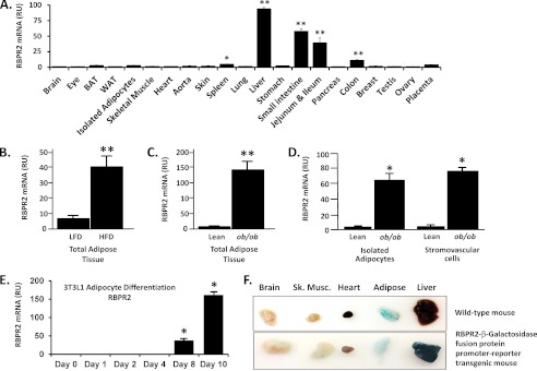
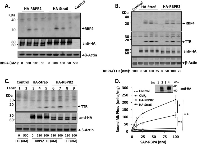
Vitamin A (retinol) is absorbed in the small intestine, stored in liver, and secreted into circulation bound to serum retinol-binding protein (RBP4). Circulating retinol may be taken up by extrahepatic tissues or recycled back to liver multiple times before it is finally metabolized or degraded. Liver exhibits high affinity binding sites for RBP4, but specific receptors have not been identified. The only known high affinity receptor for RBP4, Stra6, is not expressed in the liver. Here we report discovery of RBP4 receptor-2 (RBPR2), a novel retinol transporter expressed primarily in liver and intestine and induced in adipose tissue of obese mice. RBPR2 is structurally related to Stra6 and highly conserved in vertebrates, including humans. Expression of RBPR2 in cultured cells confers high affinity RBP4 binding and retinol transport, and RBPR2 knockdown reduces RBP4 binding/retinol transport. RBPR2 expression is suppressed by retinol and retinoic acid and correlates inversely with liver retinol stores in vivo. We conclude that RBPR2 is a novel retinol transporter that potentially regulates retinol homeostasis in liver and other tissues. In addition, expression of RBPR2 in liver and fat suggests a possible role in mediating established metabolic actions of RBP4 in those tissues.


Figures

FIGURE 1.
FIGURE 1.
Alignment of Stra6, mouse 1300002K09Rik (RBPR2), and human RBPR2 orthologue chains A and B. Novel human RBPR2 chain A and chain B sequences, Hs-RBPR2-A and Hs-RBPR2-B, were cloned by 5′-RACE and 3′-RACE from a liver cDNA library and aligned with human (Hs) Stra6, murine (Mm) Stra6, and murine (Mm) RBPR2 using ClustalW (76). 5 of 6 amino acids with documented missense mutations causing loss of Stra6 function in HMS9 are conserved in the alignment. A proposed RBP4 binding site on Stra6 is also conserved. Homology of individually aligned amino acids is rated as exact (*), conserved (:), or partially conserved (.). Overall homology between murine Stra6 and RBPR2 is 17.8% based on this alignment.
FIGURE 2.
FIGURE 2.
Conserved structural elements in human Stra6, murine 1300002K09Rik (RBPR2), and RBPR2 human orthologues. A, graphical alignment of conserved structural elements. The number of amino acids (aa) in the open reading frame encoding each protein is indicated. Solid black boxes indicate locations of conserved amino acids corresponding to Stra6 HMS9 mutations and proposed RBP4 binding domain. Shaded boxes indicate locations of possible transmembrane domains, based on predictive algorithms. Predicted transmembrane domains 10 and 11 of Stra6 (asterisks) may form part of an extended intracellular hydrophobic domain based on mapping studies by Kawaguchi et al. (17). B, alignment of RBPR2 coding exons (17 exons total) and flanking genes on mouse chromosome 4B1 with corresponding locations of RBPR2 Chain A (5 exons), RBPR2 Chain B (11 exons), and flanking genes at separate loci on human chromosome 9. The schematic illustrates only the relative positions of the exon sequences; distances between the genetic loci are not drawn to scale.
FIGURE 3.
FIGURE 3.
Subcellular localization of RBPR2. A, immunofluorescence confocal microscopy of fixed, permeabilized HEK-293 cells transfected with plasmids expressing C terminus HA-tagged RBPR2 (HA-RBPR2) or Stra6 (HA-Stra6). Top panels, anti-HA immunofluorescence; middle panels, membrane staining (endomembranes and plasma membranes) with wheat germ agglutinin-fluorescein (WGA) in green and DAPI nuclear staining in blue; bottom panels, co-localization of HA-tagged receptors with WGA-stained membranes. Increasing brightness of the pseudocolored images indicates increasing receptor-membrane co-localization. B, Western blotting of cytosol (C) and membrane (M) fractions of HEK-293 cells transfected with HA-RBPR2 (lanes 1, 2, 5, and 6) or HA-Stra6 (lanes 3, 4, 7, and 8). The indicated fractions (lanes 5–8) were treated in vitro with an endoglycosidase mix before SDS-PAGE. Western blots of the same membrane and cytosol fractions for transferrin receptor (TfR) and the IGF1 receptor (IGF1R), two known transmembrane receptors, are shown as markers for the quality of the fractions.
FIGURE 4.
FIGURE 4.
Tissue expression pattern of RBPR2. A, RBPR2 mRNA was measured in tissues of wild type FVB mice by qRT-PCR. Five different commercial primer/probe sets were tested with similar results (Solaris assay results are shown). 8–9-week-old male and female mice (n = 3 for each gender) were sacrificed in the early morning fed state. Relative expression of RBPR2 was determined separately for individual tissues and normalized to liver, which was assigned relative units (RU) equal to 100. Bars, average ± S.E. (error bars) of relative expression for six mice except for ovary, testis, and breast (n = 3 of male or female gender). Similar results were obtained using two other commercial primer/probe sets (Applied Biosystems; not shown). *, p < 0.05 versus mean relative expression level for all tissues; **, p < 0.01 versus mean relative expression level for all tissues. B, RBPR2 mRNA was measured by qRT-PCR (Solaris) in whole adipose tissue of C57Bl6 mice fed a high fat diet (HFD) or low fat control diet (LFD) for 12 weeks starting at weaning (n = 8 mice/group, males, age 15–16 weeks). **, p < 0.01 versus LFD. C, RBPR2 mRNA was measured by qRT-PCR (Solaris) in whole adipose tissue of leptin-deficient LepLep(ob/ob) mice or lean Lep(+/?) littermate controls (n = 6 mice/group, males, age 4–5 months) fed normal chow. **, p < 0.01 versus lean control. D, adipocytes and stromovascular cells were isolated by collagenase digestion and buoyancy centrifugation from perigonadal fat pads of leptin-deficient LepLep(ob/ob) mice or lean Lep(+/?) littermate controls (n = 6 mice/group, males, age 4–5 months) fed normal chow. mRNA was measured by qRT-PCR (Solaris). E, RBPR2 mRNA was measured by qRT-PCR (Applied Biosystems) in cultured 3T3L1 preadipocytes just prior to confluence (day 0) and at the indicated time points during differentiation to mature adipocytes containing multiple lipid droplets. Adipocyte differentiation was maximal by day 10 based on lipid accumulation (not shown). Three biological replicates were measured for each condition. F, expression of RBPR2-β-galactosidase fusion protein in RBPR2 promoter-reporter transgenic mice was detected by overnight staining of tissues with X-gal substrate. Blue coloration produced by X-gal cleavage indicates expressed β-galactosidase activity (top, tissues from representative C57Bl6 wild type mouse; bottom, tissues from representative littermate RBPR2 promoter-reporter mouse). A high level of endogenous, nonspecific galactosidase activity was detected in the intestine of both the wild type and transgenic promoter-reporter mice, preventing assessment of reporter expression in that tissue (not shown). A significant amount of endogenous galactosidase activity was also present in adipose tissue (top and bottom panels), and total activity did not differ significantly between wild type (top) and transgenic promoter-reporter mice (bottom).
FIGURE 5.
FIGURE 5.
RBPR2 mediates binding of RBP4 to cells. For A–C, HEK-293 cells transfected with C terminus HA-tagged RBPR2 or Stra6 (HA-RBPR2 and HA-Stra6) or empty plasmid control (pcDNA3.1), as indicated, were incubated in serum-free medium containing different purified proteins (RBP4 alone, RBP4·TTR complex, or TTR alone). Cells were washed to remove unbound proteins, lysed, and analyzed by SDS-PAGE and Western blotting. A, incubations were performed with indicated concentrations of purified human RBP4; Western blots are shown for bound RBP4 (top), transfected receptors (middle), or β-actin as a loading control (bottom). B, incubations were performed with the indicated concentrations of purified human RBP4·TTR complex; Western blots are shown for bound RBP4 (top panel), bound TTR (second panel), transfected receptors (third panel) or β-actin as a loading control (bottom panel). C, incubations were performed with the indicated concentrations of purified human TTR; Western blots are shown for bound TTR (top), transfected receptors (middle), or β-actin as a loading control (bottom). D, HEK-293 cells were transfected with HA-RBPR2, HA-Stra6, or an irrelevant receptor (leptin receptor long form, ObRb) and incubated in serum-free medium with indicated concentrations of recombinant SAP-RBP4 fusion protein. Cells were washed and lysed, and binding of SAP-RBP4 fusion protein was detected by measurement of thermostable alkaline phosphatase activity (units/mg of lysate protein), as described (16). Control-transfected cells (Control) received an empty plasmid (pcDNA3.1). *, p < 0.05; **, p < 0.01 by repeated measures ANOVA for the indicated binding curve comparisons. Inset, Western blot of HA-tagged receptors in membrane preparations of cells transfected in parallel. Error bars, S.E.
FIGURE 6.
FIGURE 6.
Characterization of RBP4 binding to receptor-transfected cells. A, HEK-293 cells transfected with C terminus HA-tagged RBPR2 (HA-RBPR2) or Stra6 (HA-Stra6) were incubated in serum-free medium containing 0.5 μm purified human RBP4. Cells were (i) washed twice with PBS and lysed; (ii) treated with 100 mm sodium bicarbonate, pH 10, for 20 min to extract peripheral membrane proteins and lysed; or (iii) incubated in bicarbonate buffer, washed twice with PBS, and lysed, as indicated. Lysates were analyzed by SDS-PAGE and Western blotting to detect bound RBP4 (arrow). B, HEK-293 cells were transfected with the indicated quantities of plasmid DNA for expression of C terminus HA-tagged RBPR2 (HA-RBPR2); the “0” HA-RBPR2 condition represents cells transfected with an empty vector control plasmid (pcDNA3.1, 5 μg). Binding was performed as described for Fig. 5 and under “Experimental Procedures” and analyzed by SDS-PAGE and Western blotting to detect bound RBP4 (arrow). C and D, HEK-293 cells were transfected with expression plasmids for C terminus HA-tagged Stra6 (HA-Stra6), a positive control for RBP4 binding, or two irrelevant HA-tagged transmembrane proteins: transforming growth factor β receptor II (TGFR2) or receptor-type protein-tyrosine phosphatase α (PTPRα). Each receptor displays an extracellular HA tag based on known topology. C, anti-HA tag Western blot of membrane (M) and cytosol (C) fractions from the transfected cells. Arrows, bands in membrane fraction corresponding to HA-Stra6 (usual mobility 70–80 kDa), HA-TGFR2 (usual mobility 60–80 kDa), and HA-PTPRα (usual mobility 95–115 kDa). D, binding of purified RBP4 to HA-receptor-transfected cells was performed as described above and analyzed by SDS-PAGE and Western blotting to detect bound RBP4 (arrow).
FIGURE 7.
FIGURE 7.
RBPR2 mediates retinol uptake. A, HEK-293 cells were transfected with C terminus HA-tagged Stra6 (left, open and closed squares) or RBPR2 (right, open and closed squares). Control cells were transfected with an empty plasmid (pcDNA3.1; left and right, open circles). Where indicated, cells were additionally co-transfected with an expression plasmid for LRAT (left and right), the intracellular enzyme that produces the retinyl ester storage form of retinol. Intact cells were incubated for 30 min at 37 ºC in serum-free medium containing purified recombinant murine RBP4 or fatty acid-free bovine serum albumin (a nonspecific ligand control) loaded with [3H]retinol. The x axis shows the concentration of either retinol-bound RBP4 or retinol-bound albumin. Cells were washed and lysed, and retinol uptake was determined by scintillation counting (expressed as fmol/mg of cell lysate protein/min). **, p < 0.01 for indicated comparisons as determined by two-way ANOVA. Inset, Western blot of HA-tagged receptors in membrane preparations of cells transfected in parallel. B, [3H]retinol uptake from holo-RBP4 or bovine serum albumin (100 nm each) was measured in transfected HEK-293 cells at different time points. The mean of four biological replicates is shown for each time point and condition. Inset, anti-HA Western blot of HA-tagged RBPR2 in membrane preparations of cells transfected in parallel. *, p < 0.05 for RBP4-ROH versus control and albumin-ROH, RBPR2 conditions.
FIGURE 8.
FIGURE 8.
Endogenous RBPR2 mediates [3H]retinol uptake and RBP4 binding in hepatoma cells. H4IIe rat hepatoma cells were treated with a siRNA pool designed to knock down RBPR2 or a non-targeting control siRNA pool (NTC). Effects of siRNAs were compared with treated control cells that did not receive siRNA (Mock). A, RBPR2 knockdown was confirmed by measuring RBPR2 mRNA levels by qRT-PCR (Solaris). **, p < 0.01 for the indicated comparison. B, binding of SAP-RBP4 at the indicated concentrations was measured in control plasmid-transfected cells and cells transfected with RBPR2 siRNA or non-targeting control siRNA, as described in the legend to Fig. 5D and under “Experimental Procedures.” **, p < 0.01 for the indicated comparison by two-way ANOVA. C, [3H]retinol uptake was measured in mock-transfected cells and cells transfected with RBPR2 siRNA or NTC siRNA, as described in the legend to Fig. 7A and under “Experimental Procedures.” *, p < 0.05 for the indicated comparison. For A, three biological replicates were measured for each condition; data are representative of two repeated experiments. For B and C, six biological replicates were measured in data combined from two repeated experiments. Error bars, S.E.
FIGURE 9.
FIGURE 9.
Liver RBPR2 expression in vivo and regulation of RBPR2 mRNA by retinol and retinoic acid. A, RBPR2 mRNA was measured by qRT-PCR targeting the exon 10/11 junction (Solaris) in the liver of wild type C57Bl6 mice (n = 7, males, age 6–7 months). Retinol and retinyl ester were measured in parallel by HPLC in aliquots of the same liver samples, and averages were correlated; R is the Pearson correlation co-efficient. A two-tailed p value was calculated. Correlation with retinol and retinyl ester was also obtained (R = 0.77, p < 0.05; data not shown) for qRT-PCR targeting the exon 1/2 junction (Applied Biosystems). B, RBPR2 mRNA was measured in Hepa1 mouse hepatoma cells treated overnight in reduced serum (0.2% FBS) medium containing the indicated concentrations of human holo-RBP4 or equimolar free retinol added directly to the medium. *, p < 0.05; **, p < 0.01 versus vehicle-treated control. C, RBPR2 mRNA was measured in Hepa1 cells treated overnight with the indicated concentrations of ATRA in medium containing normal or reduced serum (10 or 0.2% FBS, respectively). **, p < 0.01 for the indicated comparisons; three biological replicates were measured. For B and C, RBPR2 was normalized to housekeeping gene GAPDH; results without normalization were comparable (not shown). D, [3H]retinol uptake from mouse holo-RBP4 (100 nm) was determined in cells pretreated overnight with 1 μm retinol or ATRA, as described in the legend to Fig. 7A and under “Experimental Procedures.” *, p < 0.05; **, p < 0.01 versus vehicle-treated control. Six biological replicates were measured in data combined from two separate experiments. Error bars, S.E.

References

    1. Norum K. R., Blomhoff R. (1992) McCollum Award Lecture, 1992. Vitamin A absorption, transport, cellular uptake, and storage. Am. J. Clin. Nutr. 56, 735–744 - PubMed
    1. Tanumihardjo S. A. (2011) Vitamin A. Biomarkers of nutrition for development. Am. J. Clin. Nutr. 94, 658S–665S - PMC - PubMed
    1. Chambon P. (1996) A decade of molecular biology of retinoic acid receptors. FASEB J. 10, 940–954 - PubMed
    1. McGrane M. M. (2007) Vitamin A regulation of gene expression. Molecular mechanism of a prototype gene. J. Nutr. Biochem. 18, 497–508 - PubMed
    1. Mendoza-Parra M. A., Walia M., Sankar M., Gronemeyer H. (2011) Dissecting the retinoid-induced differentiation of F9 embryonal stem cells by integrative genomics. Mol. Syst. Biol. 7, 538. - PMC - PubMed

Publication types