

ZBTB7B (Th-POK) regulates the development of IL-17-producing CD1d-restricted mouse NKT cells
- PMID: 23105140
- PMCID: PMC3515659
- DOI: 10.4049/jimmunol.1201486
ZBTB7B (Th-POK) regulates the development of IL-17-producing CD1d-restricted mouse NKT cells
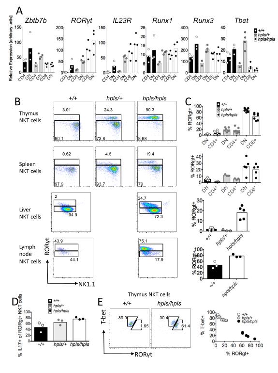
Abstract
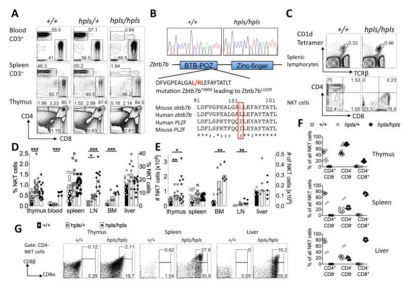
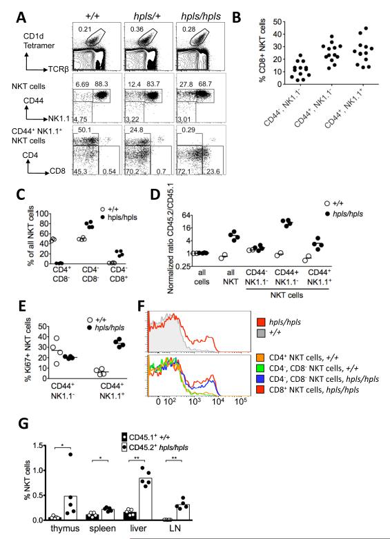
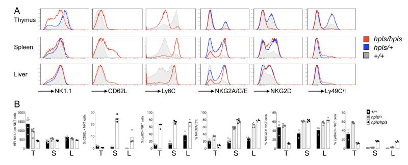
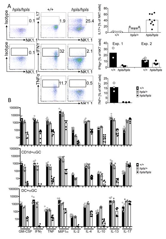
CD1d-dependent NKT cells represent a heterogeneous family of effector T cells including CD4(+)CD8(-) and CD4(-)CD8(-) subsets that respond to glycolipid Ags with rapid and potent cytokine production. NKT cell development is regulated by a unique combination of factors, however very little is known about factors that control the development of NKT subsets. In this study, we analyze a novel mouse strain (helpless) with a mis-sense mutation in the BTB-POZ domain of ZBTB7B and demonstrate that this mutation has dramatic, intrinsic effects on development of NKT cell subsets. Although NKT cell numbers are similar in Zbtb7b mutant mice, these cells are hyperproliferative and most lack CD4 and instead express CD8. Moreover, the majority of ZBTB7B mutant NKT cells in the thymus are retinoic acid-related orphan receptor γt positive, and a high frequency produce IL-17 while very few produce IFN-γ or other cytokines, sharply contrasting the profile of normal NKT cells. Mice heterozygous for the helpless mutation also have reduced numbers of CD4(+) NKT cells and increased production of IL-17 without an increase in CD8(+) cells, suggesting that ZBTB7B acts at multiple stages of NKT cell development. These results reveal ZBTB7B as a critical factor genetically predetermining the balance of effector subsets within the NKT cell population.
Figures
References
Publication types
MeSH terms
Substances
Grants and funding
LinkOut - more resources
Full Text Sources
Molecular Biology Databases
Research Materials
