

Immunomodulatory molecules are released from the first trimester and term placenta via exosomes
- PMID: 23107341
- PMCID: PMC3534832
- DOI: 10.1016/j.placenta.2012.10.005
Immunomodulatory molecules are released from the first trimester and term placenta via exosomes
Abstract
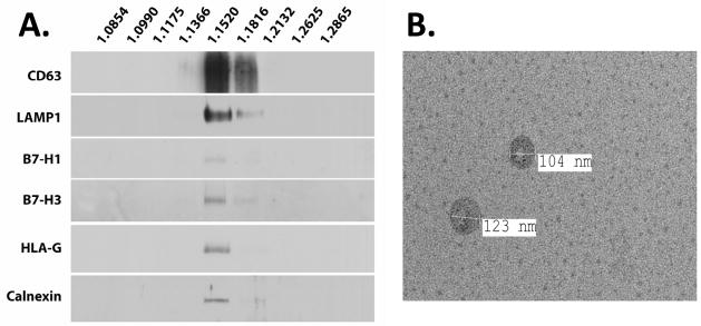
The semiallogenic fetus is tolerated by the maternal immune system through control of innate and adaptive immune responses. Trophoblast cells secrete nanometer scale membranous particles called exosomes, which have been implicated in modulation of the local and systemic maternal immune system. Here we investigate the possibility that exosomes secreted from the first trimester and term placenta carry HLA-G and B7 family immunomodulators. Confocal microscopy of placental sections revealed intracellular co-localization of B7-H1 with CD63, suggesting that B7-H1 associates with subcellular vesicles that give rise to exosomes. First trimester and term placental explants were then cultured for 24 h. B7H-1 (CD274), B7-H3 (CD276) and HLA-G5 were abundant in pelleted supernatants of these cultures that contained microparticles and exosomes; the latter, however, was observed only in first trimester pellets and was nearly undetectable in term explant-derived pellets. Further purification of exosomes by sucrose density fractionation confirmed the association of these proteins specifically with exosomes. Finally, culture of purified trophoblast cells in the presence or absence of EGF suggested that despite the absence of HLA-G5 association with term explant-derived exosomes, it is present in exosomes secreted from mononuclear cytotrophoblast cells. Further, differentiation of cytotrophoblast cells reduced the presence of HLA-G5 in secreted exosomes. Together, the results suggest that the immunomodulatory proteins HLA-G5, B7-H1 and B7-H3, are secreted from early and term placenta, and have important implications in the mechanisms by which trophoblast immunomodulators modify the maternal immunological environment.
Copyright © 2012 Elsevier Ltd. All rights reserved.
Figures


Similar articles
-
Leptin promotes HLA-G expression on placental trophoblasts via the MEK/Erk and PI3K signaling pathways.Placenta. 2015 Apr;36(4):419-26. doi: 10.1016/j.placenta.2015.01.006. Epub 2015 Jan 22. Placenta. 2015. PMID: 25649687
-
B7 family molecules are favorably positioned at the human maternal-fetal interface.Biol Reprod. 2003 May;68(5):1496-504. doi: 10.1095/biolreprod.102.010058. Epub 2002 Nov 27. Biol Reprod. 2003. PMID: 12606489
-
The immunomodulatory proteins B7-DC, B7-H2, and B7-H3 are differentially expressed across gestation in the human placenta.Am J Pathol. 2005 Aug;167(2):465-73. doi: 10.1016/S0002-9440(10)62990-2. Am J Pathol. 2005. PMID: 16049332 Free PMC article.
-
B7 family molecules: novel immunomodulators at the maternal-fetal interface.Placenta. 2002 Apr;23 Suppl A:S95-101. doi: 10.1053/plac.2002.0813. Placenta. 2002. PMID: 11978065 Review.
-
Review: Does size matter? Placental debris and the pathophysiology of pre-eclampsia.Placenta. 2012 Feb;33 Suppl:S48-54. doi: 10.1016/j.placenta.2011.12.006. Epub 2012 Jan 2. Placenta. 2012. PMID: 22217911 Review.
Cited by
-
Extracellular vesicles in host-pathogen interactions and immune regulation - exosomes as emerging actors in the immunological theater of pregnancy.Heliyon. 2019 Aug 31;5(8):e02355. doi: 10.1016/j.heliyon.2019.e02355. eCollection 2019 Aug. Heliyon. 2019. PMID: 31592031 Free PMC article. Review.
-
Trophoblast-secreted soluble-PD-L1 modulates macrophage polarization and function.J Leukoc Biol. 2020 Sep;108(3):983-998. doi: 10.1002/JLB.1A0420-012RR. Epub 2020 May 9. J Leukoc Biol. 2020. PMID: 32386458 Free PMC article.
-
Placental small extracellular vesicles: Current questions and investigative opportunities.Placenta. 2020 Dec;102:34-38. doi: 10.1016/j.placenta.2020.03.002. Epub 2020 Mar 10. Placenta. 2020. PMID: 33218576 Free PMC article. Review.
-
Regulatory T cells in embryo implantation and the immune response to pregnancy.J Clin Invest. 2018 Oct 1;128(10):4224-4235. doi: 10.1172/JCI122182. Epub 2018 Oct 1. J Clin Invest. 2018. PMID: 30272581 Free PMC article. Review.
-
Effects of miR-98 in intrauterine extracellular vesicles on maternal immune regulation during the peri-implantation period in cattle.Sci Rep. 2019 Dec 30;9(1):20330. doi: 10.1038/s41598-019-56879-w. Sci Rep. 2019. PMID: 31889113 Free PMC article.
References
-
- James E, Chai JG, Dewchand H, Macchiarulo E, Dazzi F, Simpson E. Multiparity induces priming to male-specific minor histocompatibility antigen, HY, in mice and humans. Blood. 2003;102:388–93. - PubMed
-
- Piper KP, McLarnon A, Arrazi J, Horlock C, Ainsworth J, Kilby MD, Martin WL, Moss PA. Functional HY-specific CD8+ T cells are found in a high proportion of women following pregnancy with a male fetus. Biol Reprod. 2007;76:96–101. - PubMed
-
- Verdijk RM, Kloosterman A, Pool J, van de Keur M, Naipal AM, van Halteren AG, Brand A, Mutis T, Goulmy E. Pregnancy induces minor histocompatibility antigen-specific cytotoxic T cells: implications for stem cell transplantation and immunotherapy. Blood. 2004;103:1961–4. - PubMed
Publication types
MeSH terms
Substances
Grants and funding
LinkOut - more resources
Full Text Sources
Other Literature Sources
Research Materials
Miscellaneous
