Review
. 2012 Nov;24(4):525-36.
doi: 10.1016/j.coms.2012.08.003.

Orbital anatomy for the surgeon

Affiliations
Review

Orbital anatomy for the surgeon

Timothy A Turvey et al. Oral Maxillofac Surg Clin North Am. 2012 Nov.

Abstract

An anatomic description of the orbit and its contents and the eyelids directed toward surgeons is the focus of this article. The bone and soft tissue anatomic nuances for surgery are highlighted, including a section on osteology, muscles, and the orbital suspensory system. Innervation and vascular anatomy are also addressed.


Figures

Fig. 1
Fig. 1
Avulsion of the eye occurred as a result of a deceleration injury in which the patient also sustained severe midfacial fractures. This is an example of deceleration forces exceeding the strength of the lid retractors, suspensory and check ligaments, and the natural shape of the orbit where the internal diameter exceeds the diameter of the orbital rims. (Patient treated at Parkland Memorial Hospital, Dallas, TX, under the direction of Dr R.V. Walker.)
Fig. 2
Fig. 2
The 7 bones of the orbit. (From Rougier J, Tessier P, Hervouet F, et al. Chirurgie plastique orbito-palpébrale. Paris: Elsevier Masson SAS; 1977. Copyright © Société Française d’Ophtalmologie. All rights reserved; with permission.)
Fig. 3
Fig. 3
Superior orbital fissure syndrome consists of ptosis, proptosis, pupillary dilation, and ophthalmoplegia. (A) Ptosis associated with the condition. (B) Radiograph demonstrating a fracture extending into the superior orbital fissure (arrows demonstrate orbital fracture). (C) Pupillary dilation of the right eye in another patient with superior orbital fissure syndrome. (D, E, F) Ophthalmoplegia. (Patient treated at John Peter Smith Hospital, Ft. Worth, TX, under the direction of Drs Bruce Epker and Larry Wolford.)
Fig. 4
Fig. 4
The anatomy of the medial aspect of the palpebral fissure. (Reprinted from Romanes GJ. Cunningham’s textbook of anatomy. 10th edition. Oxford Press; 1962. Fig. 957, p. 803; with permission.)
Fig. 5
Fig. 5
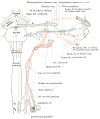
Sympathetic innervation of the orbital contents arising from the superior cervical ganglion and entering the orbit via the first division of the trigeminal nerve and the oculomotor nerve. A., artery; div., division; Inf., inferior; M., muscle; N., nerve; palp. sys., palpabrae superioris; Sup., superior; Symp., sympathetic. (Reprinted from Romanes GJ. Cunningham’s textbook of anatomy. 10th edition. Oxford Press; 1962. Fig. 56, p. 692; with permission.)
Fig. 6
Fig. 6
The orbit and eyelids. Notice the elaborate labyrinth of muscles, tendons, ligaments, and fascia, which contribute to the movement, suspension, and containment of the ocular globe. Inf., inferior; Ir, lateral rectus; ir, inferior rectus; Is, levator superiorus; lig., ligament; m., muscle; mr, medial rectus; sup., superior. (Reprinted from Manson P, Clifford CM, Su CT, et al. Mechanisms of global support and posttraumatic enophthalmos: I. The anatomy of the ligament sling and its relation to intramuscular cone orbital fat. Plast Reconstr Surg 1986;77(2):193–202. Fig. 6, p. 198; with permission from Williams Wilkins Publishing Co.)
Fig. 7
Fig. 7
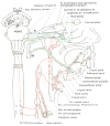
The secretory innervation of the lacrimal gland via parasympathetic fibers arising from the facial nerve ganglion. br., branch; Inf., inferior; Int., internal; N., nerve; Sup., superior. (Reprinted from Romanes GJ. Cunningham’s textbook of anatomy. 10th edition. London: Oxford Press; 1962. Fig. 868, p. 703; with permission.)
Fig. 8
Fig. 8
(A, B) Arterial blood supply of the orbit and its contents. (Reprinted from Romanes GJ. Cunningham’s textbook of anatomy. 10th edition. Oxford Press; 1962; with permission.)
Fig. 8
Fig. 8
(A, B) Arterial blood supply of the orbit and its contents. (Reprinted from Romanes GJ. Cunningham’s textbook of anatomy. 10th edition. Oxford Press; 1962; with permission.)

References

    1. Ochs MW, Buckley MJ. Anatomy of the orbit. Oral Maxillofac Surg Clin North Am. 1993;5:419–29. - PubMed
    1. Tessier P, Rougier J, Herrouat F, et al. In: Plastic surgery of the orbit and eyelids: report of the French Society of Ophthalmology. Wolfe SA, translator. New York: Masson Publishing; 1981. original work published 1977.
    1. Hollingshead WH. Anatomy for surgeons. Vol. 1. Philadelphia: Harper and Row Publishers; 1982.
    1. Romanes GJ. Cunningham’s textbook of anatomy. 10. London: Oxford University Press; 1964.
    1. Rontal E, Rontal M, Guilford FT. Surgical anatomy of the orbit. Ann Otol Rhinol Laryngol. 1979;88:382–6. - PubMed

Publication types