Multicenter Study
. 2012 Nov;135(Pt 11):3392-403.
doi: 10.1093/brain/aws231. Epub 2012 Oct 29.

Adults with RRM2B-related mitochondrial disease have distinct clinical and molecular characteristics

Affiliations
Multicenter Study

Adults with RRM2B-related mitochondrial disease have distinct clinical and molecular characteristics

Robert D S Pitceathly et al. Brain. 2012 Nov.

Abstract

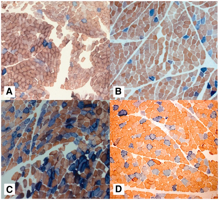
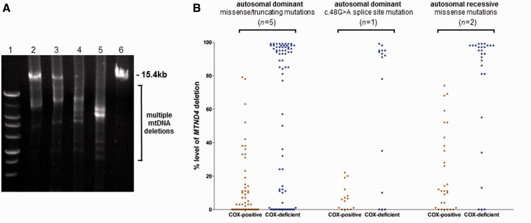
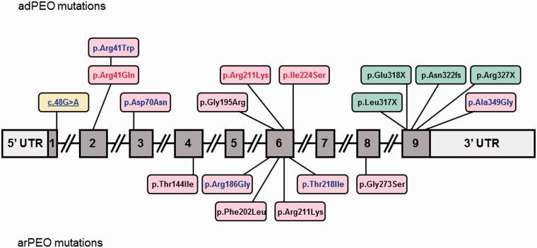
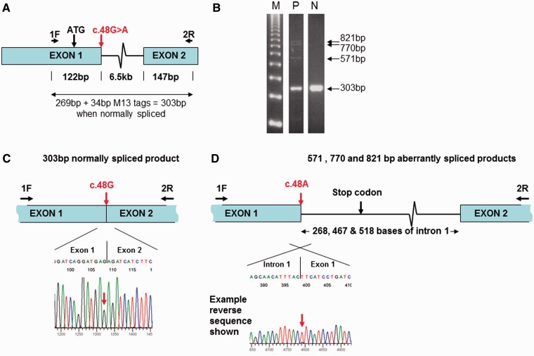
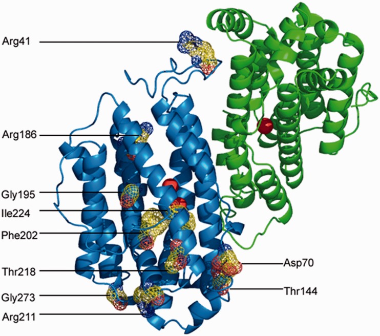
Mutations in the nuclear-encoded mitochondrial maintenance gene RRM2B are an important cause of familial mitochondrial disease in both adults and children and represent the third most common cause of multiple mitochondrial DNA deletions in adults, following POLG [polymerase (DNA directed), gamma] and PEO1 (now called C10ORF2, encoding the Twinkle helicase) mutations. However, the clinico-pathological and molecular features of adults with RRM2B-related disease have not been clearly defined. In this multicentre study of 26 adult patients from 22 independent families, including five additional cases published in the literature, we show that extra-ocular neurological complications are common in adults with genetically confirmed RRM2B mutations. We also demonstrate a clear correlation between the clinical phenotype and the underlying genetic defect. Myopathy was a prominent manifestation, followed by bulbar dysfunction and fatigue. Sensorineural hearing loss and gastrointestinal disturbance were also important findings. Severe multisystem neurological disease was associated with recessively inherited compound heterozygous mutations with a mean age of disease onset at 7 years. Dominantly inherited heterozygous mutations were associated with a milder predominantly myopathic phenotype with a later mean age of disease onset at 46 years. Skeletal muscle biopsies revealed subsarcolemmal accumulation of mitochondria and/or cytochrome c oxidase-deficient fibres. Multiple mitochondrial DNA deletions were universally present in patients who underwent a muscle biopsy. We identified 18 different heterozygous RRM2B mutations within our cohort of patients, including five novel mutations that have not previously been reported. Despite marked clinical overlap between the mitochondrial maintenance genes, key clinical features such as bulbar dysfunction, hearing loss and gastrointestinal disturbance should help prioritize genetic testing towards RRM2B analysis, and sequencing of the gene may preclude performance of a muscle biopsy.


Figures

Figure 1
Figure 1
Mitochondrial histochemical changes associated with RRM2B mutations. Representative sequential COX-SDH histochemistry demonstrates a mosaic distribution of COX-deficient muscle fibres (blue) among fibres exhibiting normal COX activity (brown). Illustrated are the images for (A) Patient 5, (B) Patient 10, (C) Patient 19 and (D) Patient 20. Patients 5 and 10 have autosomal-dominant RRM2B mutations and a milder histochemical COX defect compared with Patients 19 and 20 (autosomal-recessive RRM2B mutations), in whom a more severe biochemical defect is clearly apparent.
Figure 2
Figure 2
Characterization of multiple mitochondrial DNA deletions in muscle from patients with RRM2B mutations. (A) Representative long-range PCR amplification (15.4-kb fragment) across the major mitochondrial DNA arc shows evidence of multiple mitochondrial DNA deletions in patient muscle. Lane 1, size marker; Lane 2, Patient 5; Lane 3, Patient 10; Lane 4, Patient 19; Lane 5, Patient 20; Lane 6, Control subject. Patients with autosomal-dominant mutations (Lanes 2 and 3) show amplification of wild-type full-length mitochondrial DNA amplimers in addition to mitochondrial DNA deletions, whereas those with recessive RRM2B mutations (Patients 19 and 20) display a more severe secondary mitochondrial DNA defect. (B) Quantitative single-fibre real-time-PCR reveals the majority, but not all, of COX-deficient fibres contain high levels of a clonally expanded mitochondrial DNA deletion involving the MT-ND4 gene. Autosomal- dominant missense or truncating mutations are represented by Patients 3, 5, 9, 10 and 14. Patient 1.1 has a dominant splicing mutation, whereas Patients 19 and 20 harbour recessive RRM2B mutations.
Figure 3
Figure 3
Schematic representation of the RRM2B gene structure illustrating the 18 different mutations identified in this study. Coding exons are numbered 1–9. Missense mutations are shown in pink boxes, exon 9 truncating mutations are shown in green boxes and the c.48G>A splice mutation is shown in a yellow box. RRM2B mutations associated with autosomal-dominant PEO (adPEO) in this study, which have also been associated with more severe autosomal-recessive disease [either early-onset autosomal-recessive PEO (arPEO) or mitochondrial DNA depletion syndrome], are highlighted in red. Novel unreported RRM2B mutations are highlighted in blue.
Figure 4
Figure 4
Molecular analysis of the novel c.48G>A (p.Glu16Glu) RRM2B mutation. (A) Schematic showing the location of primers and the c.48G>A mutation within RRM2B exons 1 and 2. (B) Agarose gel electrophoresis of amplified complementary DNA from Patient 1 (P) and a normal age-matched control (N) alongside a 123-bp molecular weight marker (M), with the position of normally spliced (303 bp) and aberrantly spliced (571, 770 and 821 bp) products indicated. (C) Sequencing of the normally spliced complementary DNA product from Patient 2 reveals that this product is exclusively derived from the normal c.48G allele (equivalent results for Patients 1 and 1.1 not shown). (D) Example of a sequencing trace of the aberrantly spliced products (571-bp fragment from Patient 2 is shown), demonstrating that these are almost exclusively derived from the mutant c.48G>A allele.
Figure 5
Figure 5
Location of missense RRM2B mutations on crystal structure. An image of the p53R2 dimer structure is shown (Protein Data Bank code 3HF1). The locations of 11 of the 12 amino acids altered by missense mutations identified in this study are shown on the di-iron bound (active) subunit (it has not been possible to illustrate Ala349, as the C-terminus is absent from the crystal structure). The physical space occupied by these 11 amino acids is illustrated by a multicoloured mesh, with blue indicating positive charge, red indicating negative charge and yellow indicating neutral. Iron atoms are represented as red spheres.

References

    1. Acham-Roschitz B, Plecko B, Lindbichler F, Bittner R, Mache CJ, Sperl W, et al. A novel mutation of the RRM2B gene in an infant with early fatal encephalomyopathy, central hypomyelination, and tubulopathy. Mol Genet Metab. 2009;98:300–4. - PubMed
    1. Blakely E, He L, Gardner JL, Hudson G, Walter J, Hughes I, et al. Novel mutations in the TK2 gene associated with fatal mitochondrial DNA depletion myopathy. Neuromuscul Disord. 2008;18:557–60. - PubMed
    1. Bornstein B, Area E, Flanigan KM, Ganesh J, Jayakar P, Swoboda KJ, et al. Mitochondrial DNA depletion syndrome due to mutations in the RRM2B gene. Neuromuscul Disord. 2008;18:453–9. - PMC - PubMed
    1. Bourdon A, Minai L, Serre V, Jais JP, Sarzi E, Aubert S, et al. Mutation of RRM2B, encoding p53-controlled ribonucleotide reductase (p53R2), causes severe mitochondrial DNA depletion. Nat Genet. 2007;39:776–80. - PubMed
    1. Fratter C, Raman P, Alston CL, Blakely EL, Craig K, Smith C, et al. RRM2B mutations are frequent in familial PEO with multiple mtDNA deletions. Neurology. 2011;76:2032–4. - PMC - PubMed

Publication types

MeSH terms