. 2012 Oct 29;14(5):R233.
doi: 10.1186/ar4076.

Muscle-fiber transdifferentiation in an experimental model of respiratory chain myopathy

Muscle-fiber transdifferentiation in an experimental model of respiratory chain myopathy

Nils Venhoff et al. Arthritis Res Ther. .

Abstract

Introduction: Skeletal muscle fiber composition and muscle energetics are not static and change in muscle disease. This study was performed to determine whether a mitochondrial myopathy is associated with adjustments in skeletal muscle fiber-type composition.

Methods: Ten rats were treated with zidovudine, an antiretroviral nucleoside reverse transcriptase inhibitor that induces a myopathy by interfering with mitochondrial functions. Soleus muscles were examined after 21 weeks of treatment. Ten untreated rats served as controls.

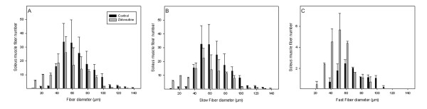
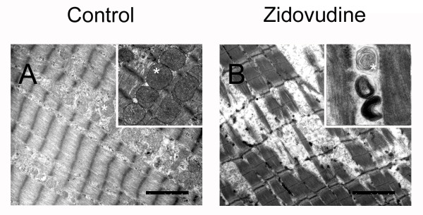
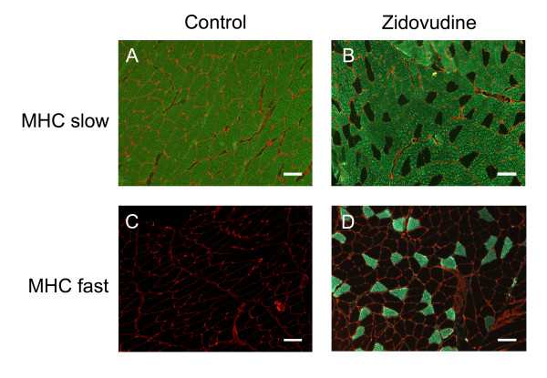
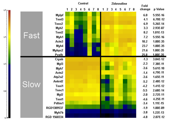
Results: Zidovudine induced a myopathy with mitochondrial DNA depletion, abnormalities in mitochondrial ultrastructure, and reduced cytochrome c oxidase activity. Mitochondrial DNA was disproportionally more diminished in type I compared with type II fibers, whereas atrophy predominated in type II fibers. Compared with those of controls, zidovudine-exposed soleus muscles contained an increased proportion (256%) of type II fibers, whereas neonatal myosin heavy chains remained repressed, indicating fiber-type transformation in the absence of regeneration. Microarray gene-expression analysis confirmed enhanced fast-fiber isoforms, repressed slow-fiber transcripts, and reduced neonatal fiber transcripts in the mitochondrial myopathy. Respiratory chain transcripts were diminished, whereas the enzymes of glycolysis and glycogenolysis were enhanced, indicating a metabolic adjustment from oxidative to glycolytic capacities. A coordinated regulation was found of transcription factors known to orchestrate type II fiber formation (upregulation of MyoD, Six1, Six2, Eya1, and Sox6, and downregulation of myogenin and ERRγ).

Conclusions: The type I to type II fiber transformation in mitochondrial myopathy implicates mitochondrial function as a new regulator of skeletal muscle fiber type.


Figures

Figure 1
Figure 1
Average numbers of soleus muscle fiber diameters (A). Fiber thinning in type I (B) and in type II fibers (C). Values represent group means (±SD).
Figure 2
Figure 2
Representative electron micrographs demonstrate zidovudine-induced degeneration of the myofibrillar lattice. Abnormal mitochondria (star) with disrupted crystal architecture. Magnification bars, 2.5 μm.
Figure 3
Figure 3
Immunohistochemistry demonstrating increased numbers of type II (fast) fibers in zidovudine-exposed soleus muscle (magnification bars, 100 μm).
Figure 4
Figure 4
Soleus muscle gene transcripts indicative of fast or slow myofiber type. The cell color codes in the heat map indicate relative transcript amounts in the soleus muscles of eight control rats and eight rats treated with zidovudine. Fast myofiber transcripts: Mylpf, myosin light chain, phosphorylatable (skeletal fast); Tnnt3, troponin T type 3 (skeletal, fast); troponin C type 2 (skeletal fast); Myh2, myosin, heavy polypeptide 2 (skeletal muscle, adult); Tnni2, troponin I type 2 (skeletal, fast); Myh1, myosin, heavy polypeptide 1 (skeletal muscle, adult); Actn3, actinin α3; Myh4, myosin, heavy-chain 4, (skeletal muscle); Mybpc2, myosin-binding protein C, fast-type; Pvalb, parvalbumin. Slow myofiber transcripts: Cryab, crystallin, αB; Myl3, myosin, light-chain 3, alkali; (ventricular, skeletal, slow); Myh7, myosin, heavy-chain 7, cardiac muscle, β; Actn2, actinin α2; Atp2a2, ATPase, Ca2+ transporting, cardiac muscle, slow twitch 2; Ankrd2, ankyrin repeat domain 23; Tnni1, troponin I type 1 (skeletal, slow); Tnnc1, troponin C type 1 (slow); Myl2, myosin, light polypeptide 2, regulatory, cardiac, slow; Tnnt1, troponin T type 1 (skeletal, slow); Pdlim1, PDZ and LIM domain 1; RGD1309537, similar to myosin regulatory light-chain 2-A, smooth muscle isoform (myosin RLC-A); Myh7b, myosin, heavy-chain 7B, cardiac muscle, β; RGD1560334, similar to myosin light-chain 1 slow α.

References

    1. Emslie-Smith AM, Engel AG. Microvascular changes in early and advanced dermatomyositis: a quantitative study. Ann Neurol. 1990;27:343–356. doi: 10.1002/ana.410270402. - DOI - PubMed
    1. Banker BQ. Dermatomyostis of childhood, ultrastructural alteratious of muscle and intramuscular blood vessels. J Neuropathol Exp Neurol. 1975;34:46–75. doi: 10.1097/00005072-197501000-00005. - DOI - PubMed
    1. Kissel JT, Halterman RK, Rammohan KW, Mendell JR. The relationship of complement-mediated microvasculopathy to the histologic features and clinical duration of disease in dermatomyositis. Arch Neurol. 1991;48:26–30. doi: 10.1001/archneur.1991.00530130034016. - DOI - PubMed
    1. Fleming JN, Schwartz SM. The pathology of scleroderma vascular disease. Rheum Dis Clin North Am. 2008;34:41–55. doi: 10.1016/j.rdc.2008.01.001. - DOI - PubMed
    1. Jobin J, Maltais F, Doyon JF, LeBlanc P, Simard PM, Simard AA, Simard C. Chronic obstructive pulmonary disease: capillarity and fiber-type characteristics of skeletal muscle. J Cardiopulmon Rehabil. 1998;18:432–437. doi: 10.1097/00008483-199811000-00005. - DOI - PubMed

Publication types

MeSH terms

Substances