. 2013 Mar;61(3):349-60.
doi: 10.1002/glia.22437. Epub 2012 Oct 25.

Toll-like receptor 4 is required for α-synuclein dependent activation of microglia and astroglia

Affiliations
Free PMC article

Toll-like receptor 4 is required for α-synuclein dependent activation of microglia and astroglia

Lisa Fellner et al. Glia. 2013 Mar.
Free PMC article

Abstract

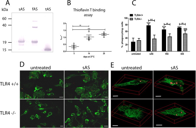
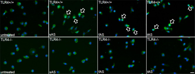
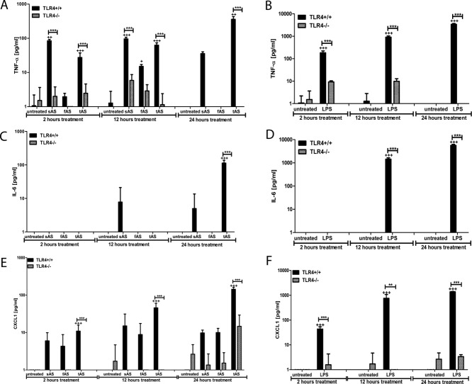
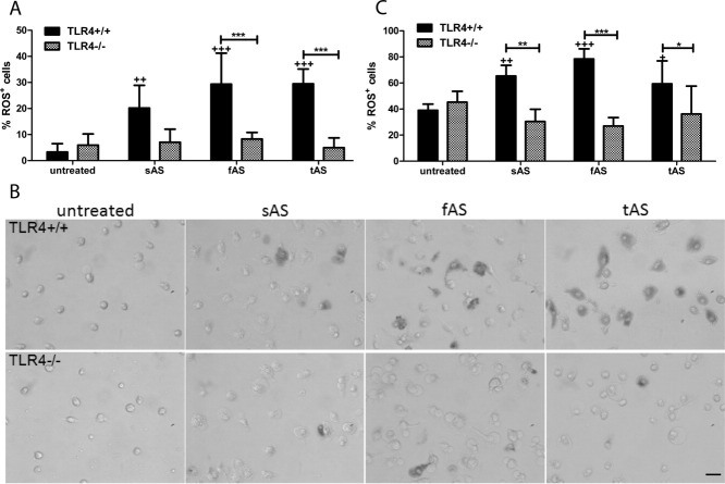
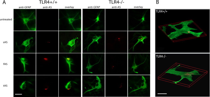
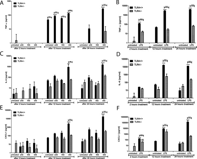
Alpha-synucleinopathies (ASP) are neurodegenerative disorders, characterized by accumulation of misfolded α-synuclein, selective neuronal loss, and extensive gliosis. It is accepted that microgliosis and astrogliosis contribute to the disease progression in ASP. Toll-like receptors (TLRs) are expressed on cells of the innate immune system, including glia, and TLR4 dysregulation may play a role in ASP pathogenesis. In this study we aimed to define the involvement of TLR4 in microglial and astroglial activation induced by different forms of α-synuclein (full length soluble, fibrillized, and C-terminally truncated). Purified primary wild type (TLR4(+/+)) and TLR4 deficient (TLR4(-/-)) murine microglial and astroglial cell cultures were treated with recombinant α-synuclein and phagocytic activity, NFκB nuclear translocation, cytokine release, and reactive oxygen species (ROS) production were measured. We show that TLR4 mediates α-synuclein-induced microglial phagocytic activity, pro-inflammatory cytokine release, and ROS production. TLR4(-/-) astroglia present a suppressed pro-inflammatory response and decreased ROS production triggered by α-synuclein treatment. However, the uptake of α-synuclein by primary astroglia is not dependent on TLR4 expression. Our results indicate the C-terminally truncated form as the most potent inductor of TLR4-dependent glial activation. The current findings suggest that TLR4 plays a modulatory role on glial pro-inflammatory responses and ROS production triggered by α-synuclein. In contrast to microglia, the uptake of alpha-synuclein by astroglia is not dependent on TLR4. Our data provide novel insights into the mechanisms of α-synuclein-induced microglial and astroglial activation which may have an impact on understanding the pathogenesis of ASP.


Figures

Figure 1
Figure 1
Microglial phagocytic activity is increased after AS-treatments, but diminished in TLR4-deficient microglia. Initially, the purity and oligomeric state of the recombinant AS preparations was determined. A: Immunoblot was performed of full length soluble, fibrillized (14-day old), and C-terminally truncated AS (sAS, fAS, and tAS). In addition to the monomer band fAS shows different oligomer species. B: The fibrillization of AS incubated at 37°C was controlled using the Thioflavin T assay. The amount of fibrillized AS was significantly augmented after 14 days at 37°C. No significant change in the degree of fibrillization was detected between AS probes after 14-day and 20-day incubation. Therefore, 14-day-old fibrillized AS (fAS) was used for all the experiments. Data were analyzed by one-way analysis of variance for multiple comparisons. ***P < 0.001, **P < 0.01 compared with 5-day incubation. Next, primary murine microglia were treated with 3 μM sAS (full length soluble AS), fAS (full length fibrillized AS) or tAS (C-terminally truncated AS), and fluorescent microspheres (red) were added. Cells were fixed with 4% paraformaldehyde and immunostained for CD11b (green). C: Morphometric analysis of CD11b-labeled microglia with incorporated microspheres revealed that AS treatment (all forms) led to a significant increase of phagocyting cells. TLR4 deficiency induced a decline of microglial phagocytic activity. Data were analyzed by two-way ANOVA with post-hoc Bonferroni test. Results are presented as mean ± S.D. +++P < 0.001 compared with untreated TLR4+/+ microglia, ***P < 0.001, **P < 0.01 comparison of TLR4+/+ to TLR4−/− microglia, $$$P < 0.001 compared with untreated TLR4−/− microglia. n = 5. D: Confocal microscopy of microglia revealed AS-dependent increase of phagocytosed microspheres by TLR4+/+ microglia (scale bar, 10 μm), whereas TLR4 ablated microglia featured reduced phagocytosis of fluorescent microspheres. Representative for all three treatments we show microglia treated with sAS and fluorescent microspheres. E: A representative confocal image stack confirmed the uptake of the microspheres (red) into CD11b-positive microglia, and further demonstrated that the amount of incorporated microspheres is decreased in TLR4−/− microglia compared with TLR4+/+ microglia (scale bar, 5 μm).
Figure 2
Figure 2
NF-κB nuclear translocation upon AS treatment in TLR4+/+ microglia. Murine primary microglia were treated with different AS-forms (full length soluble AS–sAS, fibrillized AS–fAS, and C-terminally truncated AS–tAS). Cells were fixed with 4% paraformaldehyde, immunostained for NF-κB (green) and counterstained with DAPI (blue). NF-κB translocation in TLR4+/+ microglia treated with AS was identified by co-localization of DAPI and NF-κB (see arrows). No co-localization was observed in untreated and AS treated TLR4−/− microglia (scale bar, 10 μm).
Figure 3
Figure 3
Inflammatory responses of TLR4+/+ and TLR4−/− microglia upon AS treatment. Cytokine measurements were performed using the fluorometric multiplex bead-based immunoassay (BenderSystems). Primary murine microglia (TLR4+/+ and TLR4−/−) were treated with different AS forms (full length soluble AS–sAS, fibrillized AS–fAS, and C-terminally truncated AS–tAS) or LPS as positive assay control for 2, 12, and 24 h. The supernatants were collected and used for quantifying the amount of secreted TNF-α (A, B), IL-6 (C, D) and CXCL1 (E, F). TLR4 deficiency lead to a significantly decreased TNF-α, IL-6, and CXCL1 production, especially after tAS and control LPS treatment, whereas the treatment with tAS induced a significantly elevated TNF-α, IL-6, and CXCL1 production in TLR4+/+ microglia. Data were analyzed by two-way ANOVA with post-hoc Bonferroni test. Data are the average (±SD) of three independent experiments. +++ P < 0.001, + P < 0.05 compared with untreated TLR4+/+ microglia, ***P < 0.001 comparison of TLR4+/+ to TLR4−/− microglia.
Figure 4
Figure 4
TLR4 ablation results in a reduction of ROS production by glial cells after exposure to AS. Primary murine microglial and astroglial cells were treated with different AS forms (full length soluble AS–sAS, fibrillized AS–fAS, and C-terminally truncated AS–tAS). Nitroblue tetrazolium chloride was added to visualize ROS production. Cells were fixed with 4% paraformaldehyde and ROS-positive cells were counted using an inverse microscope. A: Treatment with all AS-forms led to an enhanced ROS release by TLR4+/+ microglia. TLR4 deficient microglia showed a reduced production of ROS in comparison to TLR4+/+ microglia. Data were analyzed by two-way ANOVA with post-hoc Bonferroni test. Results are presented as mean ± S.D. +++ P < 0.001, ++ P < 0.01 compared with untreated TLR4+/+ microglia, ***P < 0.001 comparison of TLR4+/+ to TLR4−/− microglia. n = 3 (excl. untreated TLR4+/+ and TLR4−/− microglia n = 5). B: Light microscopy of microglia visualizing the difference of ROS production by TLR4+/+ and TLR4−/− microglia (scale bar, 10 μm). C: Treatment with all AS forms initiated augmented ROS production by TLR4+/+ astroglia, whereas, TLR4 ablated astroglia showed a decreased ROS production in comparison to TLR4+/+ astroglia. Data were analyzed by two-way ANOVA with post-hoc Bonferroni test. Results are presented as mean ± S.D. +++ P < 0.001, ++ P < 0.01, + P < 0.05 compared with untreated TLR4+/+ astroglia, ***P < 0.001, **P < 0.01, *P < 0.05 comparison of TLR4+/+ to TLR4−/− microglia n = 3.
Figure 5
Figure 5
Uptake of AS by aged primary murine astroglial cells. Astroglial cells were treated with different AS-forms (full length soluble AS–sAS, fibrillized AS–fAS, and C-terminally truncated AS–tAS). Cells were fixed with 4% paraformaldehyde and immunostained for GFAP (green) and AS (red). A: Both TLR4+/+ and TLR4−/− astroglial cells presented incorporated AS in the cytoplasm (scale bar 20 μm). B: Representative three-dimensionally reconstructed confocal images of GFAP-positive astroglia with incorporated sAS are shown (scale bar 10 μm).
Figure 6
Figure 6
Analysis of cytokine and chemokine release upon AS treatment of aged primary astroglia. FlowCytomix analyses of TNF-α (A, B), IL-6 (C, D), and CXCL1 (E, F) were performed using supernatants of AS- or LPS-treated primary murine astroglial cells after 2, 12, and 24 h. Especially tAS caused a significant cytokine/chemokine release in TLR4+/+ astroglia. In comparison, TLR4 ablation reduced the released cytokine amount significantly. Data were analyzed by two-way ANOVA with post-hoc Bonferroni test. Results are presented as mean ± S.D. +++ P < 0.001 compared with untreated TLR4+/+ astroglia, *** P < 0.001, ** P < 0.01 comparison of TLR4+/+ to TLR4−/− astroglia, and $$ P < 0.01 compared with untreated TLR4−/− astroglia. n = 3.

References

    1. Akira S. Toll-like receptors and innate immunity. Adv Immunol. 2001;78:1–56. - PubMed
    1. Al-Chalabi A, Durr A, Wood NW, Parkinson MH, Camuzat A, Hulot JS, Morrison KE, Renton A, Sussmuth SD, Landwehrmeyer BG, Ludolph A, Agid Y, Brice A, Leigh PN, Bensimon G. Genetic variants of the alpha-synuclein gene SNCA are associated with multiple system atrophy. PLoS One. 2009;4:e7114. - PMC - PubMed
    1. Alfonso-Loeches S, Pascual-Lucas M, Blanco AM, Sanchez-Vera I, Guerri C. Pivotal role of TLR4 receptors in alcohol-induced neuroinflammation and brain damage. J Neurosci. 2010;30:8285–8295. - PMC - PubMed
    1. Alvarez-Erviti L, Couch Y, Richardson J, Cooper JM, Wood MJ. Alpha-synuclein release by neurons activates the inflammatory response in a microglial cell line. Neurosci Res. 2011;69:337–342. - PubMed
    1. Apetri MM, Maiti NC, Zagorski MG, Carey PR, Anderson VE. Secondary structure of alpha-synuclein oligomers: Characterization by raman and atomic force microscopy. J Mol Biol. 2006;355:63–71. - PubMed

Publication types

MeSH terms