

Alzheimer disease family history impacts resting state functional connectivity
- PMID: 23109152
- PMCID: PMC3490438
- DOI: 10.1002/ana.23643
Alzheimer disease family history impacts resting state functional connectivity
Abstract
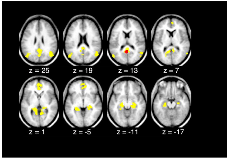
Objective: Offspring whose parents have Alzheimer disease (AD) are at increased risk for developing dementia. Patients with AD typically exhibit disruptions in the default mode network (DMN). The aim of this study was to investigate the effect of a family history of late onset AD on DMN integrity in cognitively normal individuals. In particular, we determined whether a family history effect is detectable in apolipoprotein E (APOE) ε4 allele noncarriers.
Methods: We studied a cohort of 348 cognitively normal participants with or without family history of late onset AD. DMN integrity was assessed by resting state functional connectivity magnetic resonance imaging.
Results: A family history of late onset AD was associated with reduced resting state functional connectivity between particular nodes of the DMN, namely the posterior cingulate and medial temporal cortex. The observed functional connectivity reduction was not attributable to medial temporal structural atrophy. Importantly, we detected a family history effect on DMN functional connectivity in APOE ε4 allele noncarriers.
Interpretation: Unknown genetic factors, embodied in a family history of late onset AD, may affect DMN integrity prior to cognitive impairment.
Copyright © 2012 American Neurological Association.
Figures


References
-
- Thies W, Bleiler L. 2011 Alzheimer’s disease facts and figures. Alzheimers Dement. 2011;7(2):208–44. - PubMed
-
- Gatz M, Reynolds CA, Fratiglioni L, et al. Role of genes and environments for explaining Alzheimer disease. Arch Gen Psychiatry. 2006 Feb;63(2):168–74. - PubMed
-
- Tanzi RE, Bertram L. Twenty years of the Alzheimer’s disease amyloid hypothesis: a genetic perspective. Cell. 2005 Feb 25;120(4):545–55. - PubMed
-
- Saunders AM, Strittmatter WJ, Schmechel D, et al. Association of apolipoprotein E allele epsilon 4 with late-onset familial and sporadic Alzheimer’s disease. Neurology. 1993 Aug;43(8):1467–72. - PubMed
Publication types
MeSH terms
Substances
Grants and funding
- P01AG026276/AG/NIA NIH HHS/United States
- P50 NS006833/NS/NINDS NIH HHS/United States
- R01 AG034119/AG/NIA NIH HHS/United States
- P01 AG026276/AG/NIA NIH HHS/United States
- P01AG03991/AG/NIA NIH HHS/United States
- R01AG029672/AG/NIA NIH HHS/United States
- P30 NS048056/NS/NINDS NIH HHS/United States
- R01 NR012657/NR/NINR NIH HHS/United States
- P30NS048056/NS/NINDS NIH HHS/United States
- P01AG50837/AG/NIA NIH HHS/United States
- K23MH081786/MH/NIMH NIH HHS/United States
- P01 AG003991/AG/NIA NIH HHS/United States
- R01NR012657/NR/NINR NIH HHS/United States
- P50 AG005681/AG/NIA NIH HHS/United States
- R01NR012907/NR/NINR NIH HHS/United States
- R01 AG029672/AG/NIA NIH HHS/United States
- R01 NR012907/NR/NINR NIH HHS/United States
- K23 MH081786/MH/NIMH NIH HHS/United States
- R01AG034119/AG/NIA NIH HHS/United States
- P50NS006833/NS/NINDS NIH HHS/United States
- P50AG05681/AG/NIA NIH HHS/United States
LinkOut - more resources
Full Text Sources
Medical
Miscellaneous
