

A comparison between "sandwich" and conventional methods of repairing spinal dura rupture
- PMID: 23109308
- PMCID: PMC6583603
- DOI: 10.1111/os.12005
A comparison between "sandwich" and conventional methods of repairing spinal dura rupture
Abstract
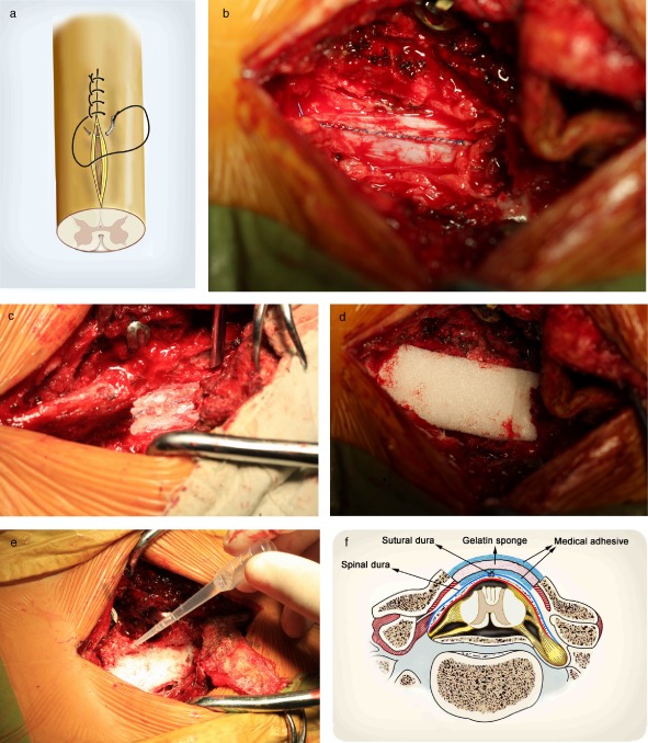
Objective: To study the therapeutic efficacy of the "sandwich" method (medical glue + gelatin sponge + medical glue) of spinal dural repair for preventing cerebrospinal fluid (CSF) leaks during treatment of subdural tumors.
Methods: Fifty-four patients with spinal subdural tumors treated between April 2007 and June 2011 were retrospectively investigated. The patients were divided into two groups: a conventional group (group A) and a "sandwich" group (group B). The group A patients included 16 males and 7 females with an average tumor course of 11 months (range, 2-34 months). Four of their 23 tumors were in the cervical spine, eight thoracic, and eleven lumbar. The group B patients included 19 males and 12 females with an average tumor course of 12 months (range, 3-36 months). Five of their 31 tumors were in the cervical spines, 10 thoracic, and 16 lumbar. In group A, the dural repairs were performed with interlocking sutures and a gelatin sponge covering the dura; whereas in group B, they were performed with interlocking sutures, painting of medical glue around the dural incision, covering this with a gelatin sponge, and finally covering of the gelatin sponge with medical glue. The total volume of drainage after operation, incidence of CSF leaks, healing of the incision, and recovery of clinical performance were recorded.
Results: Compared to group A patients, group B patients had a significantly smaller total volume of drainage (P < 0.05) on the day of surgery, and the first, second, and third postoperative days. The incidence of CSF leakage in group B was also significantly less than in group A (P < 0.05). Before discharge, three patients in group A developd hydrops, which was successfully treated by aspiration, continuous pressure from sandbags, and the prone position. During the first 3 months of follow-up, five patients developed deep hydrops under their incisions but required no treatment. There were no obvious abnormalities in group B.
Conclusion: After removal of subdural spinal tumors by incising the dura mater, or in related spinal surgery, application of the "sandwich" complex to the damaged spinal dura reduces the volume of drainage postoperatively and reduces the incidence of CSF leakage.
© 2012 Tianjin Hospital and Wiley Publishing Asia Pty Ltd.
Figures


References
-
- Epstein NE. The frequency and etiology of intraoperative dural tears in 110 predominantly geriatric patients undergoing multilevel laminectomy with noninstrumented fusions. J Spinal Disord Tech, 2007, 20: 380–386. - PubMed
-
- Joseph V, Kumar GS, Rajshekhar V. Cerebrospinal fluid leak during cervical corpectomy for ossified posterior longitudinal ligament: incidence, management, and outcome. Spine, 2009, 34: 491–494. - PubMed
-
- Stendel R, Danne M, Fiss I, et al Efficacy and safety of a collagen matrix for cranial and spinal dural reconstruction using different fixation techniques. J Neurosurg, 2008, 109: 215–221. - PubMed
-
- Sun X, Sun C, Liu X, et al The frequency and treatment of dural tears and cerebrospinal fluid leakage in 266 patients with thoracic myelopathy caused by ossification of the ligamentum flavum. Spine, 2012, 37: E702–E707. - PubMed
-
- Khan MH, Rihn J, Steele G, et al Postoperative management protocol for incidental dural tears during degenerative lumbar spine surgery: a review of 3,183 consecutive degenerative lumbar cases. Spine, 2006, 31: 2609–2613. - PubMed
Publication types
MeSH terms
Substances
LinkOut - more resources
Full Text Sources
Miscellaneous
