. 2012 Oct 29;199(3):437-51.
doi: 10.1083/jcb.201203109.

Local axonal function of STAT3 rescues axon degeneration in the pmn model of motoneuron disease

Affiliations

Local axonal function of STAT3 rescues axon degeneration in the pmn model of motoneuron disease

Bhuvaneish Thangaraj Selvaraj et al. J Cell Biol. .

Abstract

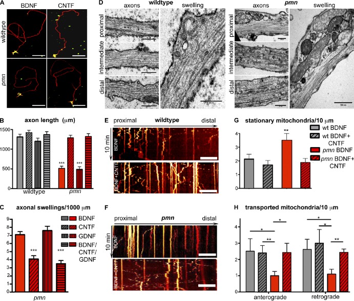
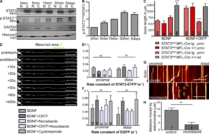
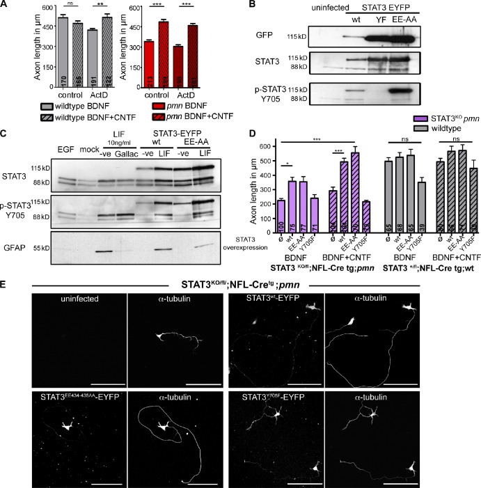
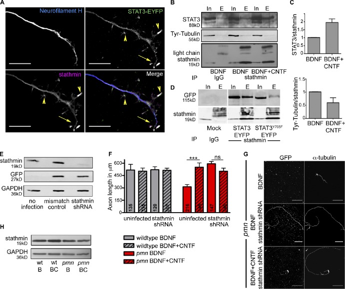
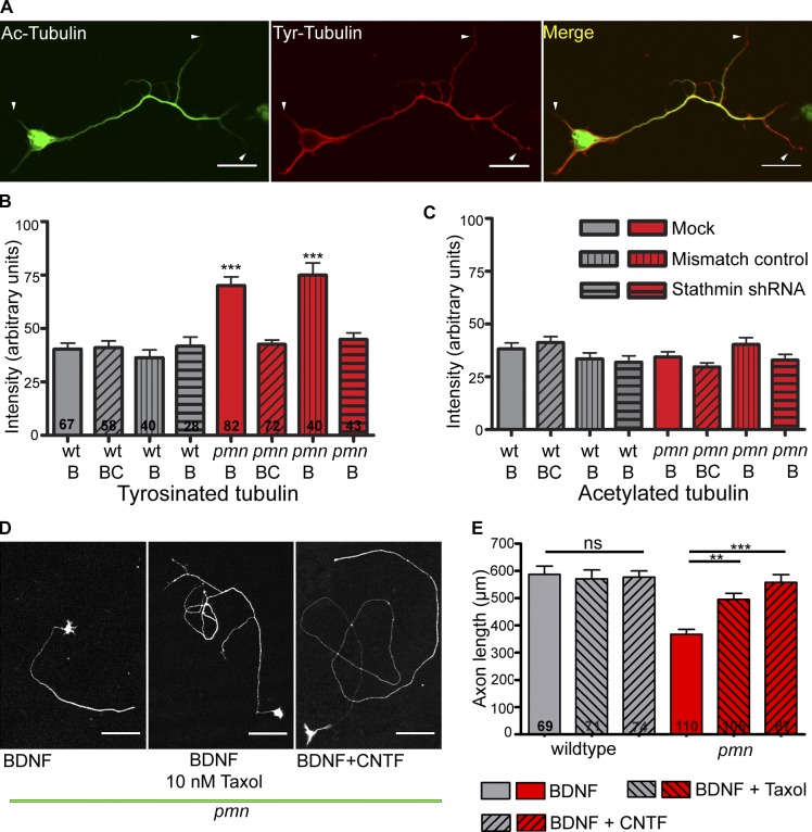
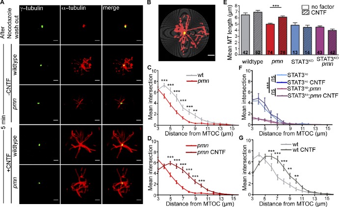
Axonal maintenance, plasticity, and regeneration are influenced by signals from neighboring cells, in particular Schwann cells of the peripheral nervous system. Schwann cells produce neurotrophic factors, but the mechanisms by which ciliary neurotrophic factor (CNTF) and other neurotrophic molecules modify the axonal cytoskeleton are not well understood. In this paper, we show that activated signal transducer and activator of transcription-3 (STAT3), an intracellular mediator of the effects of CNTF and other neurotrophic cytokines, acts locally in axons of motoneurons to modify the tubulin cytoskeleton. Specifically, we show that activated STAT3 interacted with stathmin and inhibited its microtubule-destabilizing activity. Thus, ectopic CNTF-mediated activation of STAT3 restored axon elongation and maintenance in motoneurons from progressive motor neuronopathy mutant mice, a mouse model of motoneuron disease. This mechanism could also be relevant for other neurodegenerative diseases and provide a target for new therapies for axonal degeneration.


Figures

Figure 1.
Figure 1.
CNTF rescues axon elongation and mitochondrial transport in pmn mutant motoneurons in vitro. (A) Representative images of pmn mutant and wild-type motoneurons cultured for 7 DIV in the presence of BDNF or CNTF and stained against MAP2 (green) and against tau (red). Bars, 100 µm. (B and C) Differential effect of CNTF on axon length and reduction in axonal swellings. n = 3 independent experiments. At least 50 cells were measured per condition and experiment. (D) Electron micrographs of axonal segments (left) and swellings (right) of wild-type and pmn mutant motoneurons cultured in presence of BDNF for 7 DIV, showing swellings filled with organelles in pmn mutant motoneurons. Bars: (left) 500 nm; (right) 1,000 nm. (E and F) Representative kymographs of axonal mitochondria labeled with Rhodamine 123 in wild-type and pmn mutant motoneurons cultured for 5 DIV in the presence of BDNF or BDNF and CNTF. Bars, 25 µm. (G and H) CNTF normalizes axonal transport of mitochondria in pmn mutant (54 cells for 5 ng/ml BDNF and 19 cells for 10 ng/ml BDNF + CNTF) motoneurons to levels in wild-type motoneurons (28 cells for BDNF and 12 cells for BDNF + CNTF). n = 6 independent experiments. Statistical analysis: *, P < 0.05; **, P < 0.01; ***, P < 0.001; ANOVA with Bonferroni posthoc test. wt, wild type. Data shown represent means ± SEM.
Figure 2.
Figure 2.
STAT3 is essential for CNTF-mediated axon elongation. (A) Nuclear (N) and cytoplasmic (C) fractionation followed by Western blot analysis from primary motoneurons showing activation of p-STAT3Y705 upon CNTF stimulation. Activated STAT3 is mainly found in the cytoplasm at any time point investigated. Fractionation was controlled with GAPDH (cytosolic) and histone 3 (nuclear) as markers. (B) Quantification of activated p-STAT3Y705 in the cytoplasmic fraction of motoneurons after CNTF stimulation. n = 3 independent experiments. For each individual experiment, the 0-min data point was set to 1. (C) Loss of CNTF-mediated rescue of axon length in conditional STAT3 ablated pmn mutant motoneurons. Numbers in bars indicate number of cells measured. n = 3 independent experiments. Statistical analysis: ***, P < 0.001; ANOVA with Bonferroni posthoc test. (D) Representative images of a FRAP experiment performed on STAT3-EYFP–transduced primary motoneurons cultured for 6 DIV. The bleached axonal segment is marked by the green bar in the first top image. Fluorescence recovery is depicted at various time intervals as shown. For better representation of the montage, motoneuron topology was altered using ImageJ tool straighten. Bars, 100 µm. (E and F) Diffusion rates (s−1) of STAT3-EYFP and EGFP in lentivirally transduced primary motoneurons. Diffusion rates were obtained by curve fitting using one-phase decay function and least-square fit of normalized FRAP data (see Materials and methods). Numbers in bars indicate numbers of cells analyzed. n = 3 independent experiments. P > 0.05; Kruskal-Wallis with Dunn’s multiple comparison test. (G and H) Acute treatment with 10 µM nocodazole inhibits axonal transport of MitoTracker CMXRos–labeled mitochondria. (G) Representative kymograph of nocodazole-treated motoneurons showing reduced mobility of mitochondria. (H) Quantification of the maximal distance of mitochondrial movement within 5 min. n = 3 independent experiments. Numbers in bars indicate numbers of cells analyzed. Statistical analysis: **, P < 0.01; Student’s t test. Bars, 10 µm. Data shown represent means ± SEM.
Figure 3.
Figure 3.
Transcription-independent activity of STAT3 mediates axon growth in pmn mutant motoneurons. (A) Axon length of wild-type and pmn mutant motoneurons in control or 5 nM actinomycin D–treated cultures after 5 DIV. Actinomycin D (ActD) was applied at 4 DIV for 24 h. Numbers in bars indicate numbers of cells analyzed. n = 3 independent experiments. Statistical analysis: **, P < 0.01; ***, P < 0.001; ANOVA with Bonferroni posthoc test. (B) Lentiviral overexpression of STAT3wt-EYFP, STAT3EE434–435AA-EYFP, and STAT3Y705F-EYFP in primary motoneurons. Wild-type and EE434–435AA mutant STAT3 can be activated at tyrosine 705 but not the STAT3Y705F-EYFP mutant. (C) LIF-induced GFAP induction in neural stem cells was reduced by STAT3EE434–435AA-EYFP and 10 µM galiellalactone (Gallac), indicating that mutant STAT3EE434–435AA-EYFP represses transcription of its target genes. (D) STAT3 phosphorylation, but not its transcriptional activity, is required for CNTF-mediated axon growth in pmn mutant motoneurons. Overexpression of STAT3EE434–435AA-EYFP mutant in STAT3-KO;pmn mutant motoneurons completely rescues axon growth upon CNTF application in contrast to STAT3Y705F-EYFP mutant. Ø represents uninfected, and WT, EE-AA, and Y705F represent lentiviral overexpression of STAT3WT-EYFP, STAT3EE434–435AA-EYFP, and STAT3Y705F-EYFP, respectively. Numbers in bars indicate numbers of cells analyzed. n = 3 independent experiments. Statistical analysis: *, P < 0.05; ***, P < 0.001; ANOVA with Kruskal-Wallis with Dunn’s multiple comparison test. (E) Representative images of STAT3-KO;pmn mutant motoneurons overexpressing STAT3wt-EYFP, STAT3EE434–435AA-EYFP, and STAT3Y705F-EYFP and cultured with BDNF and CNTF. Bars, 100 µm. −ve, negative; wt, wild type. Data shown represent means ± SEM.
Figure 4.
Figure 4.
Stathmin knockdown rescues axonal pathology in pmn mutant motoneurons. (A) Colocalization of STAT3-EYFP and stathmin in cultured motoneurons. Both proteins are colocalized and enriched in branch points (arrowheads) and growth cones (arrows). Bars, 10 µm. (B) Immunoprecipitation of stathmin from motoneurons cultured for 5 DIV. Western blot analysis after immunoprecipitation shows that STAT3 interaction with stathmin is enhanced after CNTF application. (first and second lanes) Input (In) and eluate (E) from IgG control; (third and fourth lanes) input and eluate from motoneurons cultured with BDNF; (fifth and sixth lanes) input and eluate from motoneurons cultured with BDNF and pulsed with CNTF for 30 min on day 5. (C) Quantification of Western blot signals shows a twofold increase in the STAT3–stathmin interaction and reduced stathmin–tyrosinated (Tyr) tubulin interaction after CNTF application; n = 3 independent experiments. (D) Immunoprecipitation experiments of stathmin with wild-type and dominant-negative STAT3 (STAT3Y705F-EYFP). Subsequent Western blot analysis shows loss of STAT3–stathmin interaction when phosphorylation at Y705 is abolished. White line indicates that intervening lanes have been spliced out. (E) Western blot analysis of protein extracts from cultured primary motoneurons after lentiviral stathmin knockdown. (F) Axon length is restored in pmn mutant motoneurons after lentiviral stathmin knockdown. Wild-type and pmn mutant motoneurons were cultured for 5 DIV. Stathmin knockdown rescues axon length in pmn mutant motoneurons. CNTF application did not induce additional axon growth in motoneurons with stathmin knockdown. Numbers in bars indicate number of cells measured. n = 3 independent experiments. ***, P < 0.001; Kruskal-Wallis with Dunn’s multiple comparison test. (G) Representative images of pmn mutant motoneurons after lentiviral stathmin knockdown. Cells were labeled with GFP and α-tubulin (Cy-3) antibodies. Bars, 100 µm. (H) CNTF application does not alter stathmin protein level in primary motoneurons of wild-type and pmn mutant mice. BDNF and CNTF are indicated by B and C, respectively. IP, immunoprecipitation; wt, wild type. Error bars represent means ± SEM.
Figure 5.
Figure 5.
MT stability is altered in pmn mutant motoneurons. (A) Wild-type motoneuron stained with antibodies against acetylated and tyrosinated α-tubulin. Acetylated (Ac) tubulin (Cy2) labels stabilized MTs and is enriched in the axons but relatively excluded from dendrites and axonal growth cones (arrowheads). Tyrosinated (Tyr) tubulin (Cy3) labels dynamic and unstable MTs, including those in dendrites and axonal tips as shown by arrowheads. Bars, 20 µm. (B and C) Levels of tyrosinated and acetylated tubulin in pmn mutant motoneurons. (B) Levels of tyrosinated tubulin were increased in pmn mutant motoneurons when compared with wild-type motoneurons. 10 ng/ml CNTF treatment or stathmin knockdown in pmn mutant motoneurons reduced tyrosinated tubulin levels to wild-type levels. (C) Levels of acetylated tubulin were unchanged under conditions investigated. Numbers in bars indicate number of cells analyzed. BDNF and CNTF are indicated by B and C, respectively. Statistical analysis: ***, P < 0.001; ANOVA with Bonferroni posthoc test. (D) Representative pictures of pmn mutant motoneurons cultured with BDNF, 10 nM taxol, and BDNF and CNTF showing increased axon length upon stabilization of MTs. Bars, 100 µm. (E) Stabilization of MTs in pmn mutant motoneurons in the presence of 10 nM taxol increased axon length in pmn motoneurons to wild-type levels. Numbers in bars represent cells measured. Statistical analysis: ***, P < 0.001; ANOVA with Bonferroni posthoc test. wt, wild type. Error bars shown represent means ± SEM from three independent experiments.
Figure 6.
Figure 6.
CNTF enhances MT regrowth in cultured motoneurons. (A) MTs were depolymerized with nocodazole, and MT regrowth was analyzed at 5 min after CNTF application in cultured wild-type and pmn mutant motoneurons. The centrosome is labeled with γ-tubulin (Cy2), and MTs were labeled with α-tubulin (Cy3). Bars, 2 µm. (B) Representative image from Sholl analysis performed on primary motoneurons with 0.25-µm step concentric circles. Bar, 2 µm. (C, D, F, and G) Graphs obtained from Sholl analysis depicting number of intersections on y axis and distance from MTOC on the x axis. (C) Comparison of MT regrowth between pmn mutant and wild-type motoneurons. (D) CNTF enhances MT regrowth in pmn mutant motoneurons. (E) Graphical representation of mean length of polymerized MTs formed in wild-type and pmn mutant motoneurons with and without CNTF and in STAT3fl/KO;NFL-Cretg motoneurons. Numbers in bars represent the number of analyzed motoneurons. Error bars shown represent means ± SEM from four independent experiments. Statistical analysis: ***, P < 0.001; ANOVA with Bonferroni posthoc test. (F) CNTF-mediated MT regrowth was abolished in STAT3fl/KO;NFL-Cretg and STAT3fl/KO;NFL-Cretg;pmn mutant motoneurons. (G) CNTF enhances MT regrowth in wild-type motoneurons. Statistical analysis: **, P < 0.01; ***, P < 0.001; two-way ANOVA with Bonferroni posthoc test. wt, wild type.

Similar articles

Cited by

References

    1. Amayed P., Pantaloni D., Carlier M.F. 2002. The effect of stathmin phosphorylation on microtubule assembly depends on tubulin critical concentration. J. Biol. Chem. 277:22718–22724 10.1074/jbc.M111605200 - DOI - PubMed
    1. Andersen J. 2003. Defects in dynein linked to motor neuron degeneration in mice. Sci. SAGE KE. 2003:PE10 10.1126/sageke.2003.18.pe10 - DOI - PubMed
    1. Bareyre F.M., Garzorz N., Lang C., Misgeld T., Büning H., Kerschensteiner M. 2011. In vivo imaging reveals a phase-specific role of STAT3 during central and peripheral nervous system axon regeneration. Proc. Natl. Acad. Sci. USA. 108:6282–6287 10.1073/pnas.1015239108 - DOI - PMC - PubMed
    1. Bender F.L., Fischer M., Funk N., Orel N., Rethwilm A., Sendtner M. 2007. High-efficiency gene transfer into cultured embryonic motoneurons using recombinant lentiviruses. Histochem. Cell Biol. 127:439–448 10.1007/s00418-006-0247-5 - DOI - PubMed
    1. Ben-Yaakov K., Dagan S.Y., Segal-Ruder Y., Shalem O., Vuppalanchi D., Willis D.E., Yudin D., Rishal I., Rother F., Bader M., et al. 2012. Axonal transcription factors signal retrogradely in lesioned peripheral nerve. EMBO J. 31:1350–1363 10.1038/emboj.2011.494 - DOI - PMC - PubMed

Publication types

MeSH terms

Substances