. 2012 Dec 1;189(11):5421-33.
doi: 10.4049/jimmunol.1200581. Epub 2012 Oct 29.

Paneth cell-mediated multiorgan dysfunction after acute kidney injury

Affiliations

Paneth cell-mediated multiorgan dysfunction after acute kidney injury

Sang Won Park et al. J Immunol. .

Abstract

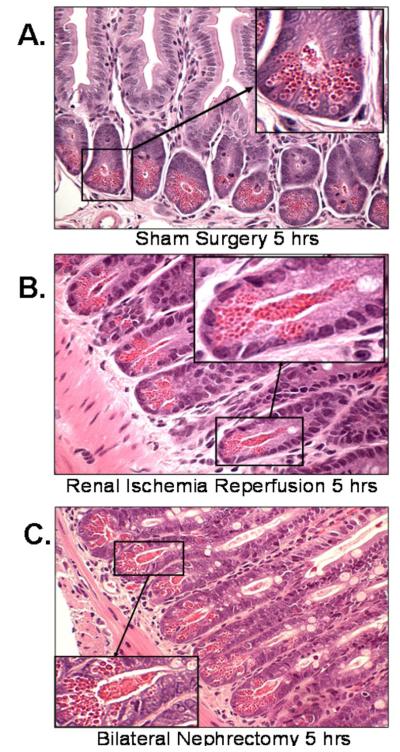
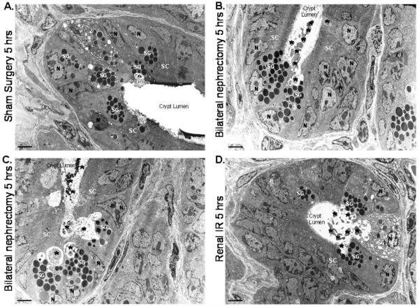
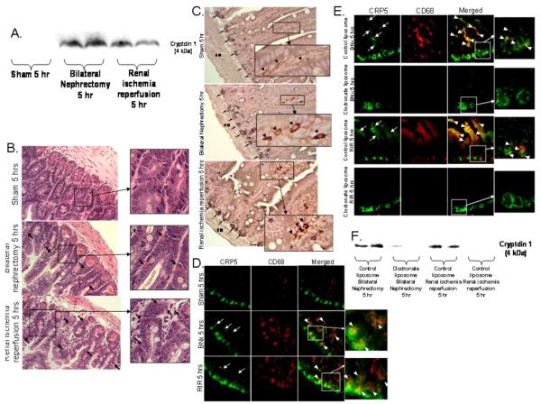
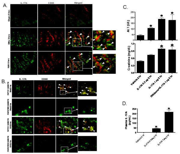
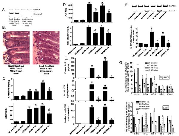
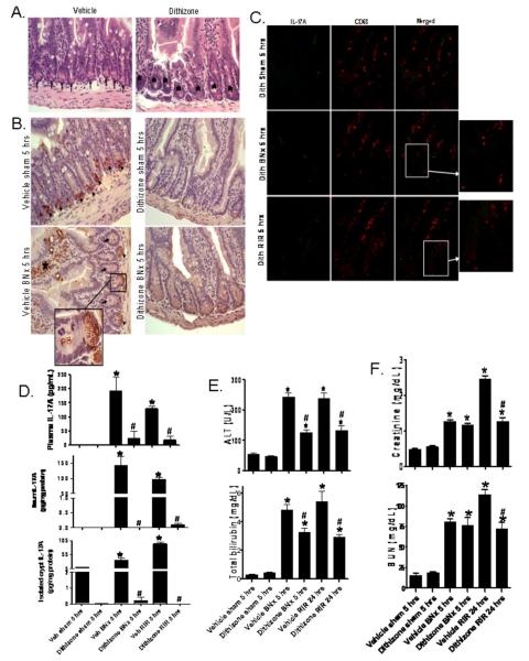
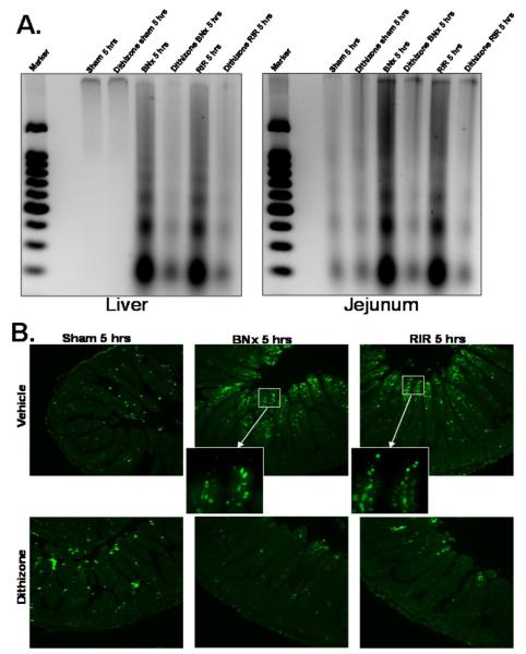
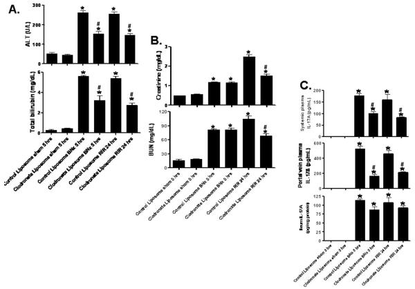
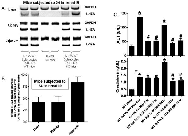
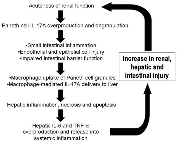
Acute kidney injury (AKI) is frequently complicated by extrarenal multiorgan injury, including intestinal and hepatic dysfunction. In this study, we hypothesized that a discrete intestinal source of proinflammatory mediators drives multiorgan injury in response to AKI. After induction of AKI in mice by renal ischemia-reperfusion or bilateral nephrectomy, small intestinal Paneth cells increased the synthesis and release of IL-17A in conjunction with severe intestinal apoptosis and inflammation. We also detected significantly increased IL-17A in portal and systemic circulation after AKI. Intestinal macrophages appear to transport released Paneth cell granule constituents induced by AKI, away from the base of the crypts into the liver. Genetic or pharmacologic depletion of Paneth cells decreased small intestinal IL-17A secretion and plasma IL-17A levels significantly and attenuated intestinal, hepatic, and renal injury after AKI. Similarly, portal delivery of IL-17A in macrophage-depleted mice decreased markedly. In addition, intestinal, hepatic, and renal injury following AKI was attenuated without affecting intestinal IL-17A generation. In conclusion, AKI induces IL-17A synthesis and secretion by Paneth cells to initiate intestinal and hepatic injury by hepatic and systemic delivery of IL-17A by macrophages. Modulation of Paneth cell dysregulation may have therapeutic implications by reducing systemic complications arising from AKI.


Figures

Figure 1
Figure 1. Paneth cell degranulation after acute kidney injury
Representative H&E staining images of small intestinal (ileum shown) Paneth cells containing dense eosinophilic granules within their apical cytoplasm (400X magnification). Both renal IR and bilateral nephrectomy resulted in small intestinal Paneth cell degranulation (B and C) in 5 hrs compared to sham-operated animals (A). Inserts show enlarged images (2000X magnification) of Paneth cells showing degranulation into the crypt lumen. Representative of 5 independent experiments.
Figure 2
Figure 2. Paneth cell degranulation after acute kidney injury observed with electron microscopy
Representative electron micrograph images (3000X magnification) of small intestinal Paneth cell degranulation (indicated by *) 5 hrs after bilateral nephrectomy (B and C) or after renal IR (D) compared to sham-operated mice (A). The crypt lumen from sham-operated mice was devoid of Paneth cell granules. Representative of 5 experiments. N = nucleus of Paneth cells. SG = secretory granules of Paneth cells. SC = stem cells located above the Paneth cells.
Figure 3
Figure 3. Paneth cell degranulation products are delivered to portal circulation via intestinal macrophage uptake
A. Acute kidney injury increases portal venous cryptdin-1. Representative (of 3 independent experiments) immunoblotting images for cryptdin-1 (4 kDa) in portal vein plasma from mice subjected to sham-operation, bilateral nephrectomy (BNx) or renal IR (RIR). Portal blood was sampled 5 hrs after surgery and processed for Tricine-SDS PAGE as described in Methods. B-E. Macrophages take up Paneth cells granules after acute kidney injury (400X magnification). B. Representative H&E stained images of small intestine of mice after sham-operation, BNx or RIR 5 hrs prior (400X magnification). Acute kidney injury results in Paneth cell degranulation and Paneth cell granule contents migrating away from the crypt base after taken up by phagocytes (arrows). Inserts show enlarged images (2000X magnification) of eosinophilic granules within phagocytes (arrows). C. Macrophages in small intestine of mice subjected to acute kidney injury (BNx or RIR) stain for both cryptdin-5 (a Paneth cell specific marker, black, arrows) and F4/80 (a macrophage specific marker, brown). Enlarged insert (2000X magnification) shows macrophages also stain for Paneth cell marker in mice subjected to BNx and renal IR (arrow heads). Representative of 5 independent experiments. SM = smooth muscle. D. Representative (of 4 independent experiments, 200X magnification and enlarged insert of 600X magnification) photographs demonstrating immunofluorescence staining for cryptdin-5 (CRP5, a Paneth cell specific marker, green) and CD68 (a macrophage specific marker, red) in small intestine of mice subjected to sham-operation (Sham), BNx or RIR. Acute kidney injury (BNx or RIR) results in Paneth cell degranulation as cryptdin-5 migrates away from the crypt base (arrows). In addition, cryptdin-5 and macrophages co-stain (yellow, arrow heads) demonstrating that degranulated cryptdin-5 is taken up by macrophages. E. Representative (of 4 independent experiments, 200X magnification and enlarged insert of 600X magnification) immunofluorescence staining for cryptdin-5 (CRP5, green) and CD68 (red) in small intestine of mice subjected to BNx or RIR with (control liposome injected) or with (clodronate liposome injected) macrophage depletion. Mice were injected with liposomes i.p. 48 hrs before sham operation or AKI. Note that cryptdin-5 no longer migrates away from the crypt base after macrophage depletion. F. Macrophage depletion with clodronate liposome decreases portal venous cryptdin-1 after AKI. Representative (of 3 independent experiments) immunoblotting images for cryptdin-1 in portal vein plasma from mice subjected to bilateral nephrectomy (BNx) or renal IR (RIR) after control liposome or clodronate liposome injection. Portal blood was sampled 5 hrs after surgery.
Figure 4
Figure 4. Intestinal macrophages take up Paneth cell-derived IL-17A to cause multi-organ dysfunction after AKI
A. Representative (of 4 independent experiments, 200X magnification and enlarged insert of 600X magnification) photographs of immunofluorescence stain for IL-17A (green) and CD68 (a macrophage specific marker, red) in small intestine of mice subjected to sham-operation (Sham), BNx or renal ischemia reperfusion (RIR). Five hrs after acute kidney injury, IL-17A immunoreactivity increased in small intestinal crypts and we show that IL-17A and CD68 co-stain (yellow, arrow heads) demonstrating that macrophages takes up IL-17A released from the Paneth cells. B. Representative (of 4 independent experiments, 200X magnification and enlarged insert of 600X magnification) immunofluorescence staining for IL-17A (green) and CD68 (red) in small intestine of mice subjected to BNx or RIR with (control liposome injected) or with (clodronate liposome injected) macrophage depletion. Mice were injected with liposomes i.p. 48 hrs before sham operation or AKI. Note that although Paneth cell IL-17A is increased after AKI in crypts after macrophage depletion, IL-17A no longer migrates away from the crypt base. C and D. Recombinant murine IL-17A (0.3 or 1 μg per mouse, iv, N=4 each) recapitulates hepatic, renal and intestinal injury in mice. C. IL-17A injection causes dose-dependent liver (ALT) and renal (creatinine) injury. IL-17A (1 μg per mouse) also caused similar degree of liver and kidney injury in mice treated with dithizone (100 mg/kg, i.v., 6 hrs prior to IL-17A injection). D. IL-17A injection (1 μg) mimics plasma IL-17A levels achieved after acute kidney injury. Plasma samples were analyzed 5 hrs after IL-17A injection. *P<0.05 vs. vehicle-treated mice.
Figure 5
Figure 5. Paneth cell deficiency with intestinal specific Sox9 deletion
Sox9 flox/flox Villin Cre+/− mice are deficient in Paneth cell marker (cryptdin-1 mRNA, A) and in Paneth cells (B, magnification 1000X) compared to wild type (Sox9 flox/flox Villin Cre−/−) mice. Arrow heads represents complete lack of Paneth cells in Sox9 flox/flox Villin Cre+/− mice. C. Paneth cell in Sox9 flox/flox Villin Cre+/− (KO) mice protects against acute kidney injury (creatinine and blood urea nitrogen) after RIR. D. Paneth cell deficiency also protects against hepatic injury (ALT and total bilirubin) after ischemic acute kidney injury (RIR) or bilateral nephrectomy (BNx) compared to Sox9 flox/flox Villin Cre−/− (WT) mice. Mice were subjected to sham-operation (Sham, N=4), BNx (N=5) or 30 min. RIR (N=5). Plasma was collected 5 hrs after BNx and 24 hrs after RIR. E. Paneth cell deficiency in Sox9 flox/flox Villin Cre+/− (KO) mice reduces plasma (N=4), small intestine (ileum shown, N=4) and isolated crypts (N=4) IL-17A levels in mice subjected to acute kidney injury (BNx and RIR). Plasma, small intestine and isolated crypt samples were analyzed 5 hrs after acute kidney injury. F. Paneth cell deficiency in Sox9 flox/flox Villin Cre+/− (KO) mice reduces IL-17A mRNA levels in isolated crypts after acute kidney injury. Small intestinal crypts were isolated 5 hrs after sham-operation (Sham, N=4), 5 hrs after BNx (N=4) or 24 hrs after 30 min. RIR (N=4). *P<0.05 vs. sham-operated mice. #P<0.05 vs. WT mice subjected to acute kidney injury. G. Paneth cell deficiency in Sox9 flox/flox Villin Cre+/− (KO) mice reduces pro-inflammatory mRNA expression (IL-17A, TNF-α, MCP-1, MIP-2 and ICAM-1) in the liver and jejunum after acute kidney injury. Tissues were extracted 5 hrs after BNx (N=4) or 24 hrs after 30 min. RIR (N=4). *P<0.05 vs. WT mice subjected to acute kidney injury. Error bars represent 1 SEM.
Figure 6
Figure 6. Paneth cell granule depletion with dithizone treatment
A. Representative H&E (of 6 experiments, magnification 400X) of ileum from mice treated with vehicle (Li2CO3) or with dithizone 6 hrs prior. Note complete depletion of Paneth cell granules (arrows) after dithizone treatment (*). B. Representative (of 5 experiments, 400X) ileum lysozyme immunostaining. Note heavy lysozyme stain in Paneth cells (arrow heads) of mice treated with vehicle (Li2CO3). Paneth cell depletion with dithizone treatment decreased lysozyme staining in Paneth cells. In addition, lysozyme staining in Paneth cells was reduced after bilateral nephrectomy (BNx) compared to sham operated mice (Sham) and we were able to detect lysozyme staining in crypt lumen (arrows and enlarged insert of 2000X magnification). Villous lysozyme staining was also evident (*). C. Representative (of 3 experiments, 200X magnification and enlarged insert, 600X magnification) immunofluorescence stain for IL-17A (green) and CD68 (red) in small intestine of mice treated with dithizone (100 mg/kg, i.v. 6 hrs prior to renal ischemia or nephrectomy) and subjected to sham-operation, BNx or renal ischemia reperfusion (RIR). Paneth cell granule depletion reduces crypt IL-17A production after AKI. D. Dithizone treatment reduces plasma IL-17A levels (analyzed 5 hrs after acute kidney injury) in mice subjected to acute kidney injury (BNx or 30 min. RIR, N=4 each). Dithizone treatment also reduced IL-17A protein upregulation in small intestine (N=4) and in isolated crypts (N=4) 5 hrs after acute kidney injury. *P<0.05 vs. sham-operated mice. #P<0.05 vs. mice subjected to vehicle treated animals subjected to acute kidney injury. Error bars represent 1 SEM. E. Paneth cell granule depletion with dithizone treatment protects against hepatic injury after ischemic acute kidney injury (RIR) or bilateral nephrectomy (BNx). Mice were subjected to sham-operation (vehicle (veh) or dithizone Sham, N=4), BNx (N=6) or 30 min. RIR (N=6). Plasma was collected 5 hrs after BNx and 24 hrs after RIR. F. Paneth cell granule depletion with dithizone treatment also protects against acute kidney injury (creatinine and blood urea nitrogen) after RIR.
Figure 7
Figure 7. Liver and small intestine (jejunum shown) apoptosis after ischemic acute kidney injury (RIR) or bilateral nephrectomy (BNx)
A. Representative gel images (of 4 experiments) demonstrating DNA laddering as an index of DNA fragmentation in the liver and jejunum from sham-operated mice (sham), mice subjected to BNx or to RIR. Tissues were harvested 5 hrs after surgery. Paneth cell granule depletion with dithizone treatment reduces liver and small intestine (jejunum shown) apoptosis after RIR or BNx. Apoptotic DNA fragments were extracted according to the methods of Herrmann et al. (29). This method of DNA extraction selectively isolates apoptotic, fragmented DNA and leaves behind the intact chromatin. B. Representative photomicrographs of TUNEL staining in small intestine (jejunum, magnification 100X) sections. Six hrs after vehicle (Li2CO3) or dithizone treatment, mice were subjected to sham operation (sham), to BNx or to RIR. Enlarged inserts (600X magnification) show that majority of TUNEL positive cells are villous capillary endothelial cells. Dithizone treatment did not induce Paneth cell or crypt apoptosis. Photographs are representative of 4 independent experiments.
Figure 8
Figure 8. Macrophage depletion with clodronate liposome
Mice were subjected to sham-operation (N=4), bilateral nephrectomy (BNx, N=5) or ischemic acute kidney injury (RIR, N=5) after control liposome or clodronate liposome injection. Mice were injected with liposomes i.p. 48 hrs before sham operation or AKI. Plasma was collected 5 hrs after BNx and 24 hrs after RIR. A. Macrophage depletion protects against hepatic injury (ALT and bilirubin) after RIR or BNx. B. Macrophage depletion also protects against acute kidney injury (creatinine and blood urea nitrogen) after RIR. C. Macrophage depletion reduces systemic and portal vein plasma IL-17A levels without decreasing ileum IL-17A levels in mice subjected to acute kidney injury (BNx or 30 min. RIR, N=4 each, analyzed 5 hrs after acute kidney injury). *P<0.05 vs. sham-operated mice. #P<0.05 vs. control liposome injected mice subjected to acute kidney injury. Error bars represent 1 SEM.
Figure 9
Figure 9. Successful reconstitution of IL-17A in IL-17A deficient mice
A. Detection of IL-17A mRNA by RT-PCR in the liver, kidney and small intestines of IL-17A deficient mice after IL-17A wild type splenocyte injection (representative of 4 experiments). Mice were subjected to renal ischemia and 24 hrs reperfusion. B. Detection of IL-17A protein with ELISA in the liver, kidney and small jejunum of IL-17A deficient mice after IL-17A wild type splenocyte injection (N=4). Mice were subjected to renal ischemia and 24 hrs reperfusion. C. Splenocyte IL-17A does not contribute to remote organ injury after AKI. IL-17A deficient mice transfused with wild type splenocytes were still protected against hepatic and renal injury after ischemic AKI (RIR) and bilateral nephrectomy (BNx). Mice were subjected to sham-operation (Sham, N=4), BNx (N=5) or 30 min. RIR (N=5). Plasma was collected 5 hrs after BNx and 24 hrs after RIR. *P<0.05 vs. sham-operated mice. #P<0.05 vs. mice subjected to acute kidney injury after transfused with wild type splenocytes.
Figure 10
Figure 10. Proposed mechanisms of acute kidney injury induced liver dysfunction and systemic inflammation
Acute loss of renal function causes small intestinal Paneth cell generation of IL-17A and Paneth cell degranulation. We propos that IL-17A released by Paneth cells directly cause intestinal injury. Our data suggest that intestinal macrophages uptake of released Paneth cell granules promote portal delivery of IL-17A. This leads to hepatic injury (necrosis, inflammation and apoptosis) and increased generation and systemic release of TNF-α and IL-6 propagating multi-organ injury and systemic inflammation. The mechanisms that cause Paneth cells to produce increased IL-17A and degranulate after AKI remain to be determined.

References

    1. Chertow GM, Burdick E, Honour M, Bonventre JV, Bates DW. Acute kidney injury, mortality, length of stay, and costs in hospitalized patients. J. Am. Soc. Nephrol. 2005;16:3365. - PubMed
    1. Jones DR, Lee HT. Perioperative renal protection. Best. Pract. Res. Clin. Anaesthesiol. 2008;22:193. - PubMed
    1. Elapavaluru S, Kellum JA. Why do patients die of acute kidney injury? Acta Clin. Belg. 2007;(Suppl):326. - PubMed
    1. Paladino JD, Hotchkiss JR, Rabb H. Acute kidney injury and lung dysfunction: a paradigm for remote organ effects of kidney disease? Microvasc. Res. 2009;77:8. - PMC - PubMed
    1. Grigoryev DN, Liu M, Hassoun HT, Cheadle C, Barnes KC, Rabb H. The local and systemic inflammatory transcriptome after acute kidney injury. J. Am. Soc. Nephrol. 2008;19:547. - PMC - PubMed

Publication types

MeSH terms