Case Reports
. 2012;39(5):750-5.

Apical hypertrophic cardiomyopathy: preliminary attempt at palliation with use of subselective alcohol ablation

Affiliations
Case Reports

Apical hypertrophic cardiomyopathy: preliminary attempt at palliation with use of subselective alcohol ablation

Paolo Angelini et al. Tex Heart Inst J. 2012.

Abstract

We report a case of severe apical hypertrophic cardiomyopathy in order to discuss the nature of this unusual condition and the possibility of using selective alcohol ablation to effectively treat symptomatic hypertrophic cardiomyopathy that presents with apical aneurysm. A 73-year-old woman with severe, progressive dyspnea and intermittent chest pain was found to have localized left ventricular apical dyskinesia distal to an obstructive mid-distal muscular ring. The ring caused total systolic obliteration of the apical left ventricular cavity. Apical cavity pressure was extremely high, up to 330 mmHg-200 mmHg above that in the main left ventricular cavity. Because of the danger of apical rupture and clot formation, we attempted the experimental use of alcohol ablation for effective palliation. We present our pilot experience, offer a novel interpretation of the nature of this obscure entity, and possibly justify a new catheter treatment. In addition, we discuss the developmental, pathophysiologic, and clinical implications of this unusual form of hypertrophic cardiomyopathy. To our knowledge, ours is the first reported use of subselective, modified-protocol alcohol septal ablation to treat an obstructive mid-apical muscular ring in a patient with apical hypertrophic cardiomyopathy.

Keywords: Cardiomyopathy, hypertrophic/complications/epidemiology/physiopathology/therapy; ethanol/administration & dosage/therapeutic use; heart septum/pathology; hypertrophy, left ventricular/diagnosis; myocardial ischemia/complications; treatment outcome.


Figures

None
Fig. 1 Electrocardiographic tracing on admission shows ST-segment changes and deep negative T waves in the anterior leads, with signs of left ventricular hypertrophy.
None
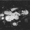
Fig. 4 Magnetic resonance imaging frames (cross-sectional views at the mid-cavity obstructive ring). A) Preoperative diastolic, B) systolic, and C) postoperative diastolic, and D) systolic images show that the mid-ventricular ring opening was essentially obliterated in both preoperative and postoperative systolic times.
None
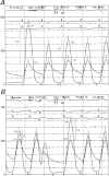
Fig. 5 Pressure tracings of the patient's states: A) preoperative and B) postoperative. A 4F pigtail catheter was placed in the apical cavity and a 6F guiding catheter was placed in the aorta. The recorded gradients varied during the procedure. The peak systolic gradient was 230 mmHg after a premature ventricular contraction and 125 mmHg during regular rhythm; postoperatively, they were 116 and 35 mmHg, respectively.
None
Fig. 6 Transthoracic contrast echocardiogram shows the protruding, hypertrophic obstructive ring at the mid-apical segment of the left ventricle. The apical aneurysm “filled” in early systole and emptied in early diastole, with no flow during most of systole. The completely occlusive myocardial ring provoked systolic expansion at the apex. Real-time motion image is available at www.texasheart.org/journal.
None
Fig. 7 Echocardiographic and continuous-wave Doppler images at the obstructive apical ring show a short interval of early systolic blood flow (A), which was interrupted for most of systole and followed by a higher blood-flow velocity peak in early diastole (B), at the time of the delayed diastolic relaxation. See also Figure 3. Therefore, the intraventricular systolic gradient did not seem to be caused by blood flow, but rather by systolic contraction of the obstructive ring in the presence of systolic obliteration of the apical cavity.
None
Fig. 2 Preoperative frame from the simultaneous apical ventricular and coronary imaging used during the alcohol ablation procedure shows the relationship between the obstructive apical hypertrophic ridge and the related coronary branches (arrows). A left ventricular 4F pigtail catheter was used for periodic angiographic testing and for apical pressure monitoring.
None
Fig. 3 Pressure tracings were obtained before alcohol ablation, one at the apex and one at the outflow portion of the left ventricle (and then superimposed by the timing of the QRS interval). The apical systolic pressure was approximately 250 mmHg, whereas the outflow pressure was 125 mmHg. In addition, the drop in end-systolic pressure was delayed in the apical portion by 5 to 10 ms with respect to the outflow chamber, during which time the Doppler signal indicated runoff from this cavity (as shown in Fig. 5).
None
Fig. 8 Postoperative magnetic resonance sequence shows a 2-cm-thick circumferential scar at the core of the circular obstructive apical ring. The apical cavity (A) was essentially unchanged in size and had a persistently thinned apical free wall. B = inflow of the main left ventricular cavity
None
Fig. 9 Schematic representation of the essential features of obstructive apical hypertrophic cardiomyopathy. The inflow/outflow/main cavity and the apical cavity are separated by a hypertrophic obstructive ring in diastole, which causes total obliteration of this segment during early systole. The asterisks indicate the papillary muscles that in this condition are found implanted on the obstructive ring. The obstructive mid-apical ventricular ring can be thought of as an abnormal, hypertrophic apical plane made of dense, coalescent trabeculations, which results in an intrinsic mechanism of progressive apical cavity enlargement and thinning of the apical free wall. Alleviation of the apical hypertension can be obtained by eliminating the mid-ventricular obstruction, by impairing the contractility of (or ablating) the myocardial hypertrophic ring, or both of these.

Comment in

References

    1. Maron MS, Olivotto I, Zenovich AG, Link MS, Pandian NG, Kuvin JT, et al. Hypertrophic cardiomyopathy is predominantly a disease of left ventricular outflow tract obstruction. Circulation 2006;114(21):2232–9. - PubMed
    1. Angelini P. The “1st septal unit” in hypertrophic obstructive cardiomyopathy: a newly recognized anatomo-functional entity, identified during recent alcohol septal ablation experience. Tex Heart Inst J 2007;34(3):336–46. - PMC - PubMed
    1. Maron BJ. Controversies in cardiovascular medicine. Surgical myectomy remains the primary treatment option for severely symptomatic patients with obstructive hypertrophic cardiomyopathy. Circulation 2007;116(2):196–206. - PubMed
    1. Sigwart U. Non-surgical myocardial reduction for hypertrophic obstructive cardiomyopathy. Lancet 1995;346(8969):211–4. - PubMed
    1. Lakkis NM, Nagueh SF, Kleiman NS, Killip D, He ZX, Verani MS, et al. Echocardiography-guided ethanol septal reduction for hypertrophic obstructive cardiomyopathy. Circulation 1998;98(17):1750–5. - PubMed