. 2012;8(10):e1003023.
doi: 10.1371/journal.pgen.1003023. Epub 2012 Oct 18.

Viral evasion of a bacterial suicide system by RNA-based molecular mimicry enables infectious altruism

Affiliations

Viral evasion of a bacterial suicide system by RNA-based molecular mimicry enables infectious altruism

Tim R Blower et al. PLoS Genet. 2012.

Abstract

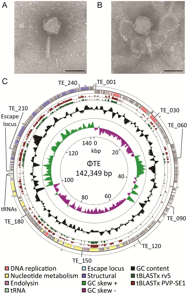
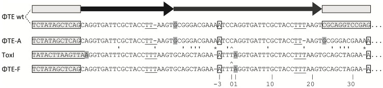
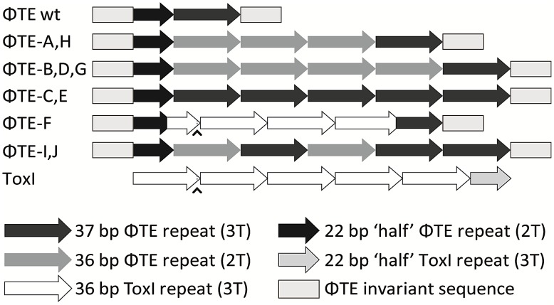
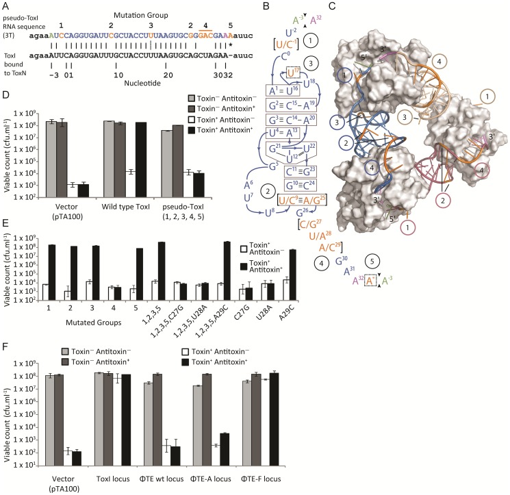
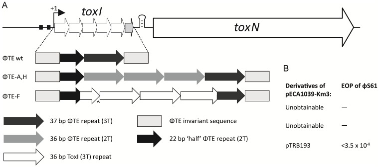
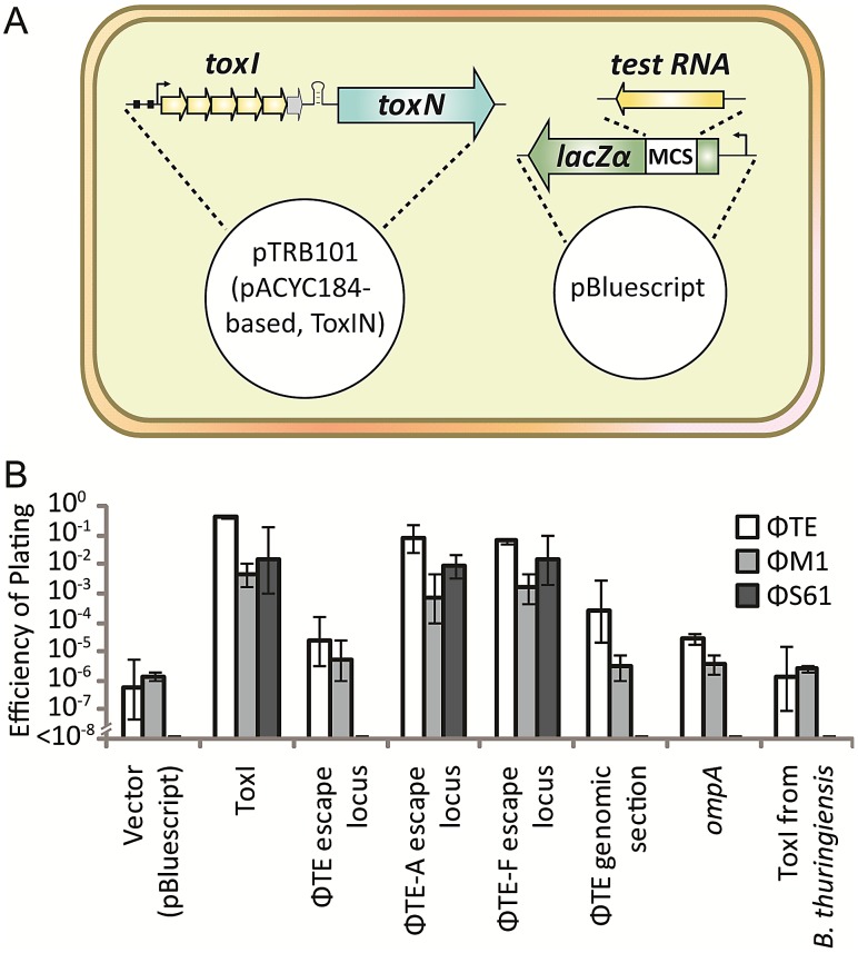
Abortive infection, during which an infected bacterial cell commits altruistic suicide to destroy the replicating bacteriophage and protect the clonal population, can be mediated by toxin-antitoxin systems such as the Type III protein-RNA toxin-antitoxin system, ToxIN. A flagellum-dependent bacteriophage of the Myoviridae, ΦTE, evolved rare mutants that "escaped" ToxIN-mediated abortive infection within Pectobacterium atrosepticum. Wild-type ΦTE encoded a short sequence similar to the repetitive nucleotide sequence of the RNA antitoxin, ToxI, from ToxIN. The ΦTE escape mutants had expanded the number of these "pseudo-ToxI" genetic repeats and, in one case, an escape phage had "hijacked" ToxI from the plasmid-borne toxIN locus, through recombination. Expression of the pseudo-ToxI repeats during ΦTE infection allowed the phage to replicate, unaffected by ToxIN, through RNA-based molecular mimicry. This is the first example of a non-coding RNA encoded by a phage that evolves by selective expansion and recombination to enable viral suppression of a defensive bacterial suicide system. Furthermore, the ΦTE escape phages had evolved enhanced capacity to transduce replicons expressing ToxIN, demonstrating virus-mediated horizontal transfer of genetic altruism.


Conflict of interest statement

The authors have declared that no competing interests exist.

Figures

Figure 1
Figure 1. ΦTE morphology and genome overview.
(A–B) Transmission electron micrographs of individual ΦTE virus particles. The tail is fully extended in (A) and contracted in (B). Each scale bar represents 100 nm. (C) Summary of the 142,349 bp circularly-permuted genome of ΦTE, including all ORFs (colour coded to function where possible), two tRNAs and the ncRNA comprising pseudo-ToxI, encoded by the escape locus (Table S2). Selected ΦTE genes are indicated by “TE_x” around the genome, for orientation. GC skew and GC content are shown along with the tBLASTx results against two related phages, coliphage rv5 and Salmonella phage PVP-SE1.
Figure 2
Figure 2. DNA alignment of ΦTE-phage escape loci and comparison with ToxI.
Long grey boxes enclose invariant sequences bordering the pseudo-ToxI repeats. Grey shaded bases indicate the start of a DNA repeat. Single, boxed, bases mark the start of a ToxI antitoxic RNA and the predicted equivalent pseudo-ToxI RNAs in the ΦTE phages. Prime symbols denote single base-pair differences between sequences. Asterisks indicate the single base addition at the end of each pseudo-ToxI RNA repeat. A circumflex, ∧, indicates the single variable base in ToxI, where the first repeat shows a T in this position rather than a C for all other repeats. The numbering system identifies the 36 nucleotides within an antitoxic ToxI pseudoknot, numbered according to position relative to the DNA repeat, thereby beginning at base −3, −2, −1, 0 and through to base 32 . Underlined bases denote the variable 2T or 3T sequences.
Figure 3
Figure 3. Schematic of the ΦTE-phage escape loci and ToxI.
Each escape phage has expanded the number of DNA repeats, whilst ΦTE-F has recombined with toxI. The first ToxI repeat encoded by ΦTE-F matches the first ToxI repeat of pECA1039, as can be seen by the presence of the non-consensus T (∧) and dotted lines. Full sequences are in Table S3.
Figure 4
Figure 4. Analysis of pseudo-ToxI as a potential antitoxin.
(A) Alignment of the pseudo-ToxI and ToxI RNA sequences. Pseudo-ToxI nucleotides are coloured to match (B) and (C), with the green and purple bases denoting the 5′ and 3′ ends of a single pseudoknot, respectively. Mutated nucleotides in pseudo-ToxI are coloured orange and numbered according to their grouping, whilst the asterisk indicates the additional 3′ nucleotide. The dotted line connecting the U in group 3 indicates the uracil that is deleted in the case of expanded repeats with 2T sequences rather than 3T. (B) Schematic of the ToxI pseudoknot. Each position containing a mutation in the pseudo-ToxI RNA has been bracketed, with the ToxI base separated from the pseudo-ToxI base by a ‘/’. The mutations have been grouped 1–5, according to position, and highlighted in orange, with the 5′ and 3′ termini in green and violet, respectively. Indels, such as U17 that is deleted in some pseudo-ToxI repeats, and the additional A* inserted in all, have been bordered by a dashed line. Base interactions are indicated by black lines, and duplex and triplex base-interactions are bordered in grey. (C) Detail of the ToxIN trimer with each pseudoknot shown either in blue, purple or beige. Each ToxN monomer is shown as a grey surface. The blue pseudoknot is oriented relative to (B). The positions of mutation groups are shown, with the group number encircled in the same colour as the corresponding pseudoknot. The additional nucleotide of group 5 is not visible as this was not in the original solved ToxIN structure. PDB: 2XDB. (D) Pseudo-ToxI cannot protect from ToxN in an over-expression assay. Protection assays were conducted as per Materials and Methods using strains of E. coli DH5α carrying both pTA49 (inducible ToxN) and a second inducible antitoxin vector as shown, including use of pTA100 as a vector-only control, “vector”. Error bars indicate the standard deviation of triplicate data. (E) Protection assays using mutants of ToxI carried out as in (D) with the antitoxin mutations in each construct numbered as per (B). (F) Protection assays carried out as in (D), testing the full escape loci of ΦTE wt, ΦTE-A and ΦTE-F with full ToxI as a positive control. Under these conditions, there was sufficient antitoxin present to inhibit induced ToxN even without specific induction of the ToxI and ΦTE-F constructs.
Figure 5
Figure 5. Only the recombinant ΦTE-F escape locus can replace ToxI in the native ToxIN locus.
(A) Organisation of the ToxIN operon. Promoter elements are shown as black boxes. The transcriptional start site is indicated by the arrow with ‘+1’. The 5.5 repeats of toxI are followed by a stem-loop terminator structure, then toxN. It was possible to excise the full toxI sequence and then attempt to replace it with the escape locus sequences, including the invariant ends, from ΦTE wt, ΦTE-A and ΦTE-F, as shown in Figure 2. (B) The single plasmid that could be successfully generated, which included the insert from ΦTE-F, was tested for Abi activity against ΦS61 and seen to be highly active.
Figure 6
Figure 6. An excess of pseudo-ToxI inhibits abortive infection.
(A) Strains of Pba ToxIN (pTRB101) were tested for their ability to abort infection in the presence of a second, pBluescript II SK- based, antitoxic plasmid. Putatively antitoxic “test RNA” sequences were cloned under the control of the constitutive lacZα promoter, to allow for constant, high-level, expression. (B) EOPs of ΦTE, ΦM1 and ΦS61 on double strains of Pba, as per key, using Pba ToxIN-FS (pTRB102, pBluescript II SK-) as the control strain. Inserts in the second, antitoxic, plasmids are indicated by the horizontal axis labels. Plasmid pBluescript II SK- was used as the no insert, “vector”, control. “ΦTE escape locus” includes the escape locus from wild type ΦTE, whilst the “ΦTE genomic section” is a 269 bp region of the ΦTE genome, taken several kb from the escape locus as a negative control. Error bars indicate the standard deviation of triplicate (minimum) data.
Figure 7
Figure 7. ΦTE-F expresses ToxI RNA during infection.
(A) Upper panel; an S1-nuclease protection assay was used to detect ToxI levels from a ToxIN plasmid during ΦTE wt infection, using an antisense probe against the full 5.5 repeat ToxI sequence . The antisense ToxI-probe was first hybridised to 10 µg of total RNA prepared from Pba ToxIN (pMJ4) at different times after ΦTE infection, and then followed by S1-nuclease treatment. Numbers (+) indicate the time (min) after infection or (−) without the addition of phage. Pba ToxIN-FS (pTA47) and Pba serve as positive and negative controls, respectively. A non-hybridized S1-digested probe (+S1) serves as a further negative control. DH5α 1.5 repeats (pTA96), a non-S1 digested probe (−S1) and an in vitro transcribed Hammerhead ribozyme (HHRz), which cleaves itself during transcription, serve as size markers. HHRz was prepared as described previously . Lower panel; Western blot targeting C-terminal FLAG tagged ToxN contained within total protein harvested from Pba ToxIN (pMJ4) at different time points, with (+, left) and without (−, right) phage infection. Time 0 indicates a sample taken immediately after infection. Total protein from Pba ToxIN (pMJ4) (−) serves as positive control. (B) Infection with escape phage ΦTE-F. Levels of ToxI were determined by S1-assay (upper) as described in (A) with and without infection. ToxN levels were estimated by Western blotting (lower) as described in (A). (C) Expression of the ΦTE-F ToxI locus. An S1-nuclease assay targeting ToxI was performed on total RNA of Pba (pBR322) at different times during ΦTE-F infection. Pba ToxIN (pMJ4) and DH5α serve as positive and negative controls, respectively.

Comment in

References

    1. Fozo EM, Makarova KS, Shabalina SA, Yutin N, Koonin EV, et al. (2010) Abundance of type I toxin-antitoxin systems in bacteria: searches for new candidates and discovery of novel families. Nucl Acids Res 38: 3743–3759. - PMC - PubMed
    1. Leplae R, Geeraerts D, Hallez R, Guglielmini J, Dreze P, et al. (2011) Diversity of bacterial type II toxin-antitoxin systems: a comprehensive search and functional analysis of novel families. Nucl Acids Res 39: 5513–5525. - PMC - PubMed
    1. Blower TR, Short FL, Rao F, Mizuguchi K, Pei XY, et al. (2012) Identification and classification of bacterial Type III toxin-antitoxin systems encoded in chromosomal and plasmid genomes. Nucl Acids Res doi:10.1093/nar/gks231 - DOI - PMC - PubMed
    1. Blower TR, Salmond GP, Luisi BF (2011) Balancing at survival's edge: the structure and adaptive benefits of prokaryotic toxin-antitoxin partners. Curr Opin Struc Biol 21: 109–118. - PubMed
    1. Ogura T, Hiraga S (1983) Mini-F plasmid genes that couple host cell division to plasmid proliferation. Proc Natl Acad Sci U S A 80: 4784–4788. - PMC - PubMed

Publication types

MeSH terms