Endoscopic ultrasound-guided fine needle aspiration cytology diagnosis of solid pseudopapillary neoplasm: three case reports with review of literature
- PMID: 23110037
- PMCID: PMC3479817
- DOI: 10.4132/KoreanJPathol.2012.46.4.399
Endoscopic ultrasound-guided fine needle aspiration cytology diagnosis of solid pseudopapillary neoplasm: three case reports with review of literature
Abstract
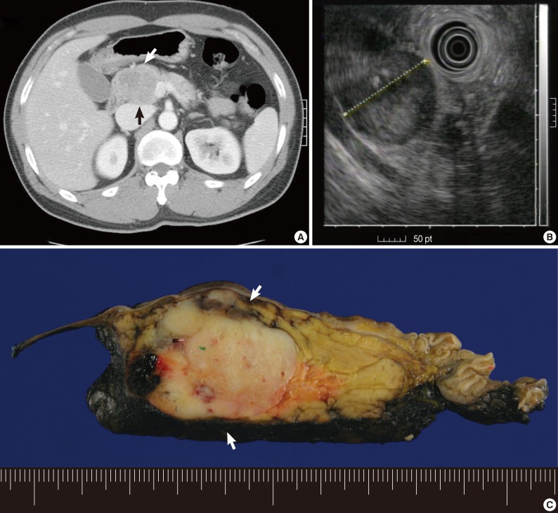
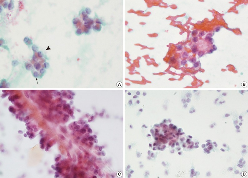
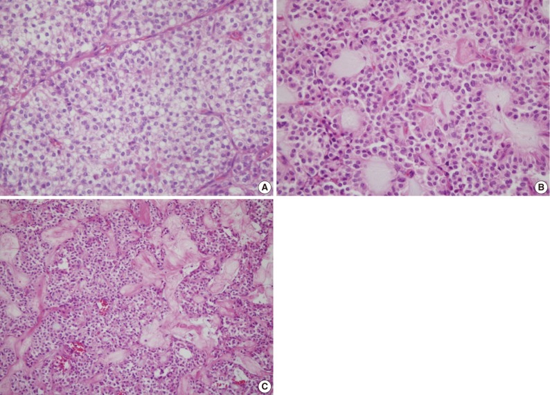
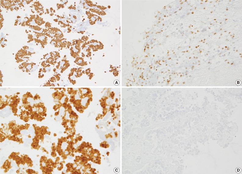
Solid pseudopapillary neoplasm of the pancreas (SPN) is relatively rare and it occurs almost exclusively in women. We recently experienced three cases of SPN diagnosed by endoscopic ultrasound-guided fine-needle aspiration (EUS-FNA). These three cases were two male and one female patient whose age was 29, 37, and 44 years old. Radiological diagnosis was pancreatic endocrine tumor (PEN) showing solid with a heterogenous echogenicity. EUS-FNA cytology specimens consisted of single cells and aggregates of uniform cells, forming microadenoid structures, branching, papillary clusters with delicate fibrovascular cores. In conclusion, a single diagnosis of SPN based on clinical and radiological findings would be risky because there is a possibility of it being misdiagnosed as PEN or other malignancies. An EUS-FNA is therefore essential for establishing the diagnosis. In addition, the pathologists should recognize the characteristic cytologic findings with immunoprofiles of SPN to prevent misdiagnosis of SPN.
Keywords: Cytology; Endoscopic ultrasound-guided fine needle aspiration; Solid pseudopapillary neoplasm.
Conflict of interest statement
No potential conflict of interest relevant to this article was reported.
Figures
References
-
- Kloppel G, Luttges J, Klimstra DS, et al. Solid pseudopapillary neoplasm. In: Hmailton SR, Aaltonene LA, editors. World Health Organization classfication of tumors: pathology and genetics of tumours of the digestive sytstem. Lyon: IARC Press; 2000. pp. 473–480.
-
- Adamthwaite JA, Verbeke CS, Stringer MD, Guillou PJ, Menon KV. Solid pseudopapillary tumour of the pancreas: diverse presentation, outcome and histology. JOP. 2006;7:635–642. - PubMed
-
- Ky A, Shilyansky J, Gerstle J, et al. Experience with papillary and solid epithelial neoplasms of the pancreas in children. J Pediatr Surg. 1998;33:42–44. - PubMed
-
- Papavramidis T, Papavramidis S. Solid pseudopapillary tumors of the pancreas: review of 718 patients reported in English literature. J Am Coll Surg. 2005;200:965–972. - PubMed
-
- Bardales RH, Centeno B, Mallery JS, et al. Endoscopic ultrasound-guided fine-needle aspiration cytology diagnosis of solid-pseudopapillary tumor of the pancreas: a rare neoplasm of elusive origin but characteristic cytomorphologic features. Am J Clin Pathol. 2004;121:654–662. - PubMed
LinkOut - more resources
Full Text Sources