. 2012;7(10):e45003.
doi: 10.1371/journal.pone.0045003. Epub 2012 Oct 22.

Tat-dependent translocation of an F420-binding protein of Mycobacterium tuberculosis

Affiliations

Tat-dependent translocation of an F420-binding protein of Mycobacterium tuberculosis

Ghader Bashiri et al. PLoS One. 2012.

Abstract

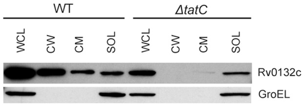
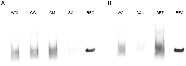
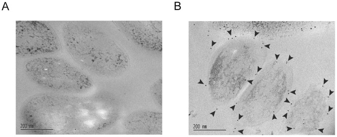
F(420) is a unique cofactor present in a restricted range of microorganisms, including mycobacteria. It has been proposed that F(420) has an important role in the oxidoreductive reactions of Mycobacterium tuberculosis, possibly associated with anaerobic survival and persistence. The protein encoded by Rv0132c has a predicted N-terminal signal sequence and is annotated as an F(420)-dependent glucose-6-phosphate dehydrogenase. Here we show that Rv0132c protein does not have the annotated activity. It does, however, co-purify with F(420) during expression experiments in M. smegmatis. We also show that the Rv0132c-F(420) complex is a substrate for the Tat pathway, which mediates translocation of the complex across the cytoplasmic membrane, where Rv0132c is anchored to the cell envelope. This is the first report of any F(420)-binding protein being a substrate for the Tat pathway and of the presence of F(420) outside of the cytosol in any F(420)-producing microorganism. The Rv0132c protein and its Tat export sequence are essentially invariant in the Mycobacterium tuberculosis complex. Taken together, these results show that current understanding of F(420) biology in mycobacteria should be expanded to include activities occurring in the extra-cytoplasmic cell envelope.


Conflict of interest statement

Competing Interests: The authors have declared that no competing interests exist.

Figures

Figure 1
Figure 1. Molecular structure of cofactor F420.
(A) Schematic representation of cofactor F420, where n varies from 2–9 in different microorganisms. (B) Mass spectrometry analysis of cofactor F420 bound to the purified Rv0132c–Δ38 protein showing the population of species differing in the number of glutamate residues in the poly-Glu tail.
Figure 2
Figure 2. SDS–PAGE and UV–visible spectra of the purified Rv0132c–Δ38 protein.
(A) SDS–PAGE gel of the purified Rv0132c–Δ38 protein. (B) UV-visible spectra for purified Rv0132c (0.5 mg/mL in PBS, red), F420 extracted from M. smegmatis cells (50 µM in PBS, green) and F420 extracted from the purified Rv0132c (blue). M: molecular weight markers (kDa).
Figure 3
Figure 3. Functional assay of Rv0132c–Δ38 protein.
The FGD activity was assessed for Rv0132c–Δ38 protein and Mtb–FGD1 as a positive control. Mtb–FGD1 shows a decrease at 420 nm absorbance (green and red lines), whereas Rv0132c–Δ38 protein indicated no change in the absorbance (yellow and blue lines). The same results were observed using various concentrations of Rv0132c–Δ38 protein in the presence of different concentrations of glucose-6-phpsphate. The graph shows assays containing 1 µM of each enzyme, 25 µM F420 with 0.1 mM (green and yellow lines) and 1 mM (red and blue lines) glucose-6-phosphate.
Figure 4
Figure 4. Structural comparison of Rv0132c with FGD1.
(A) Amino acid sequence alignment. The secondary structure elements for FGD1 are shown above the sequence. FGD1 residues that hydrogen bond with F420 or the phosphate group of glucose-6-phosphate are indicated below the sequence by F and asterisk, respectively. The twin arginines in the Tat motif and the critical cysteine residue in the lipobox motif are shown in red in the Rv0132c signal sequence. (B) Superposition of the FGD1 (orange) crystal structure on the modeled Rv0132c (cyan). The F420 cofactor (green) bound to FGD1 is shown in stick representation. Replacement of helix α9 with a smaller loop extends the active site cavity in Rv0132c. For details of FGD1 structure see .
Figure 5
Figure 5. Rv0132c export is Tat dependent.
Equalized whole cell lysates (WCL) from wild type (WT) and ΔtatC M. smegmatis expressing Rv0132c-HA were fractionated to generate cell wall (CW), cytoplasmic membrane (CM), and soluble (SOL) fractions. Fractions were separated by SDS-PAGE and proteins were detected with an anti-HA antibody. Native GroEL was detected as a cytoplasmic control. Rv0132c-HA was exported to the CW and CM fractions in wild type M. smegmatis, but it was not exported in the absence of a functional Tat pathway.
Figure 6
Figure 6. Subcellular localization of Rv0132c protein.
(A) Western blots of M. tuberculosis H37Rv subcellular fractions using 1/25000 dilution of anti–Rv0132c antiserum. Clear signals are found for the WCL, CW and CM fractions, but not for the SOL fraction. (B) Western blots of Triton X–114 treated fractions. The signal is present only in the DET fraction. WCL: whole cell lysate; CW: cell wall; CM: cytoplasmic membrane; SOL: soluble; AQU: aqueous fraction from Triton X–114 treatment; DET: detergent–enriched fraction from Triton X–114 treatment. In both panels recombinant Rv0132c–Δ38 protein (REC) is used as a positive control (0.7 µg).
Figure 7
Figure 7. Immunoelectron microscopy of the M. tuberculosis H37Ra cells using anti–Rv0132c antiserum.
Electron micrographs are shown in which thin cryo–sectioned Mtb cells are (A) treated with preimmune serum, and (B) treated with anti-Rv0132c antisera at a dilution of 1/200. The gold particles (indicated by arrowheads) are present mainly on the periphery of the cells in panel B, but are absent from the control panel (A).

References

    1. Global Tuberculosis Control (2010) World Health Organization
    1. Duncan K, Barry CE III (2004) Prospects for new antitubercular drugs. Curr Opin Microbiol 7: 460–465. - PubMed
    1. Selengut JD, Haft DH (2010) Unexpected Abundance of Coenzyme F420-dependent enzymes in the Genomes of Mycobacterium tuberculosis and other Actinobacteria. J Bacteriol 192: 5788–5798. - PMC - PubMed
    1. Bashiri G, Rehan AM, Greenwood DR, Dickson JMJ, Baker EN (2010) Metabolic engineering of cofactor F420 production in Mycobacterium smegmatis . PLoS ONE 5: e15803. - PMC - PubMed
    1. Bashiri G, Squire CJ, Moreland NJ, Baker EN (2008) Crystal structures of F420-dependent glucose-6-phosphate dehydrogenase FGD1 involved in the activation of the anti-tuberculosis drug candidate PA-824 reveal the basis of coenzyme and substrate binding. J Biol Chem 283: 17531–17541. - PubMed

Publication types

MeSH terms