. 2012;7(10):e48110.
doi: 10.1371/journal.pone.0048110. Epub 2012 Oct 26.

Natural transmission of Plasmodium berghei exacerbates chronic tuberculosis in an experimental co-infection model

Affiliations

Natural transmission of Plasmodium berghei exacerbates chronic tuberculosis in an experimental co-infection model

Ann-Kristin Mueller et al. PLoS One. 2012.

Abstract

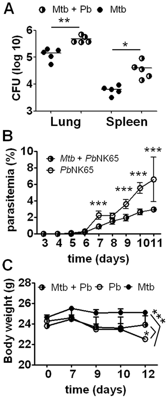
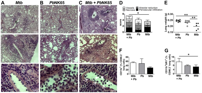
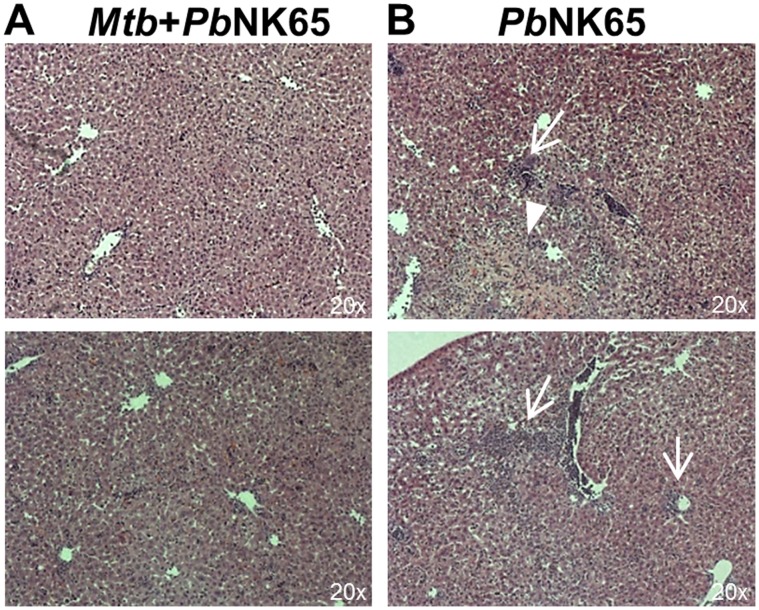
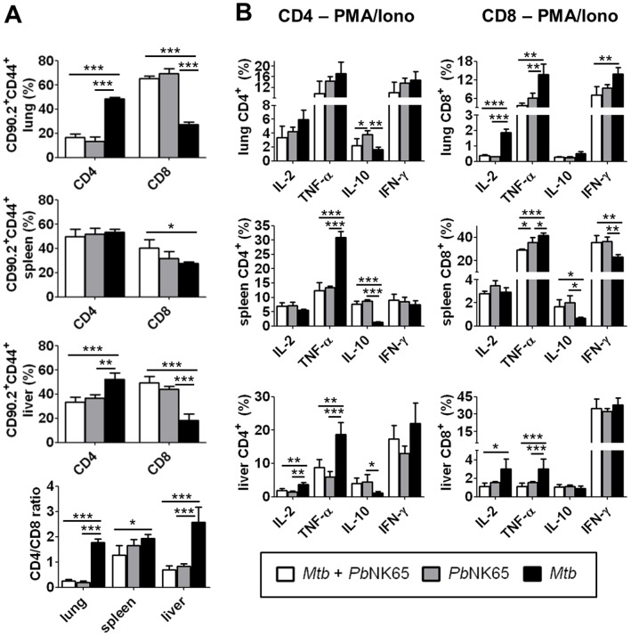
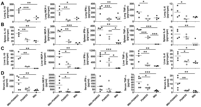
Human populations are rarely exposed to one pathogen alone. Particularly in high incidence regions such as sub-Saharan Africa, concurrent infections with more than one pathogen represent a widely underappreciated public health problem. Two of the world's most notorious killers, malaria and tuberculosis, are co-endemic in impoverished populations in the tropics. However, interactions between both infections in a co-infected individual have not been studied in detail. Both pathogens have a major impact on the lung as the prime target organ for aerogenic Mycobacterium tuberculosis and the site for one of the main complications in severe malaria, malaria-associated acute respiratory distress syndrome (MA-ARDS). In order to study the ramifications caused by both infections within the same host we established an experimental mouse model of co-infection between Mycobacterium tuberculosis and Plasmodium berghei NK65, a recently described model for MA-ARDS. Our study provides evidence that malaria-induced immune responses impair host resistance to Mycobacterium tuberculosis. Using the natural routes of infection, we observed that co-infection exacerbated chronic tuberculosis while rendering mice less refractory to Plasmodium. Co-infected animals presented with enhanced inflammatory immune responses as reflected by exacerbated leukocyte infiltrates, tissue pathology and hypercytokinemia accompanied by altered T-cell responses. Our results--demonstrating striking changes in the immune regulation by co-infection with Plasmodium and Mycobacterium--are highly relevant for the medical management of both infections in humans.


Conflict of interest statement

Competing Interests: The authors have declared that no competing interests exist.

Figures

Figure 1
Figure 1. Co-infected mice are more refractory to Plasmodium sporozoite infection but exacerbate tuberculosis.
C57BL/6 mice were aerosol infected with M. tuberculosis H37Rv (100 CFU/lung) and 40 days later challenged with PbNK65 sporozoites by mosquito bite. Control mice were infected with M. tuberculosis or PbNK65 alone, respectively. A) Mice were sacrificed 12 days after co-infection and serial dilutions of lung and spleen lysates were plated for CFU determination. B) Parasitemia in peripheral blood was monitored by daily Giemsa-stained blood smears. Note, that co-infected animals had significantly lower parasite numbers than mice infected with PbNK65 alone. Results are shown as means ± SD (n = 10). C) Weight loss was reduced when mice were pre-infected with M. tuberculosis before PbNK65 challenge. Results are shown as means ± SD (n = 10). Results from one representative experiment out of two independent ones are shown. Statistical analysis was performed using the Student’s t test (A) or ANOVA (B and C) (*p<0.05; **p<0.01; *** p<0.001).
Figure 2
Figure 2. Malaria co-infection increases inflammatory tissue responses in M. tuberculosis infected lungs.
C57BL/6 mice were aerosol infected with M. tuberculosis H37Rv (100 CFU/lung) and 40 days later challenged with PbNK65 sporozoites by mosquito bite. Control mice were infected with M. tuberculosis or PbNK65 alone, respectively. A–C) Representative H&E stains of lungs 13 days after co-infection. Note, that PbNK65 co-infection exacerbated tissue pathology compared to lungs of mice infected with M. tuberculosis alone (v  =  vessel, b  =  bronchus; asterisks indicate hemozoin loaded cells; arrow: neutrophils; arrowhead: monocytes). D) Histopathological scores from co-infected, PbNK65 or M. tuberculosis infected lungs are shown. Pathology was most severe in co-infected animals, with the total score being significantly increased compared to M. tuberculosis infected lungs (n = 4 for co-infected and PbNK65 infected; n = 5 for M. tuberculosis infected). E) Lung weights 12 days after co-infection. F–G) Lung leukocytes were analyzed for surface expression of CD11b and GR-1. Results are shown as means ± SD (n = 3–5). Results from one representative experiment out of two independent ones are shown. Statistical analysis was performed by ANOVA (*p<0.05; **p<0.01).
Figure 3
Figure 3. PbNK65 associated liver damage is reduced in co-infected mice.
C57BL/6 mice were aerosol infected with M. tuberculosis H37Rv (100 CFU/lung) and 40 days later challenged with PbNK65 sporozoites by mosquito bite. Control mice were infected with M. tuberculosis or PbNK65 alone, respectively. Representative H&E stains of liver sections 13 days after co-infection. Note, that PbNK65 infection caused periportal inflammation (B; arrows) and tissue necrosis (B; arrowhead) which was reduced in livers of co-infected animals (A).
Figure 4
Figure 4. PbNK65 co-infection alters T cell responses in M. tuberculosis infected mice.
C57BL/6 mice were infected by aerosol with M. tuberculosis H37Rv (100 CFU/lung) and challenged with PbNK65 sporozoites by mosquito bite 40 days later. Control mice were infected with M. tuberculosis or PbNK65 alone, respectively. A) 12 days upon PbNK65 infection, lungs, spleens and livers were analyzed for the presence of CD44 positive CD4 and CD8 effector T cells by flow cytometry. B) Whole lung and spleen lysates and purified liver lymphocytes were re-stimulated in vitro with PMA/Iono (50 ng/ml, respectively) and analyzed by flow cytometry for the presence of IL-2, TNF-α, IL-10 or IFN-γ producing CD4 and CD8 T cells. Results are shown as means ± SD (n = 3–5). Data from one out of two independent experiments are shown. Statistical analysis was performed by ANOVA (*p<0.05; **p<0.01; *** p<0.001).
Figure 5
Figure 5. Co-infection with M. tuberculosis and PbNK65 induces a cytokine storm.
C57BL/6 mice were infected by aerosol with M. tuberculosis H37Rv (100 CFU/lung) and challenged with PbNK65 sporozoites by mosquito bite 40 days later. Control mice were infected with M. tuberculosis or PbNK65 alone, respectively. Cytokine levels were measured in lungs (A), spleens (B), livers (C) and sera (D) 13–14 days after co-infection. Statistical analysis was performed by ANOVA and Tukey’s Multiple Comparison test. (*p<0.05; **p<0.01; ***p<0.001). Data from one out of two independent experiments are shown.

References

    1. Cooper AM (2009) Cell-mediated immune responses in tuberculosis. Annu Rev Immunol 27: 393–422. - PMC - PubMed
    1. Wolf AJ, Desvignes L, Linas B, Banaiee N, Tamura T, et al. (2008) Initiation of the adaptive immune response to Mycobacterium tuberculosis depends on antigen production in the local lymph node, not the lungs. J Exp Med 205: 105–115. - PMC - PubMed
    1. Lin PL, Plessner HL, Voitenok NN, Flynn JL (2007) Tumor necrosis factor and tuberculosis. J Investig Dermatol Symp Proc 12: 22–25. - PubMed
    1. Ulrichs T, Kaufmann SH (2006) New insights into the function of granulomas in human tuberculosis. J Pathol 208: 261–269. - PubMed
    1. Murray CJ, Rosenfeld LC, Lim SS, Andrews KG, Foreman KJ, et al. (2012) Global malaria mortality between 1980 and 2010: a systematic analysis. Lancet 379: 413–431. - PubMed

Publication types

MeSH terms