. 2012;7(10):e48310.
doi: 10.1371/journal.pone.0048310. Epub 2012 Oct 26.

Autoinducers act as biological timers in Vibrio harveyi

Affiliations

Autoinducers act as biological timers in Vibrio harveyi

Claudia Anetzberger et al. PLoS One. 2012.

Abstract

Quorum sensing regulates cell density-dependent phenotypes and involves the synthesis, excretion and detection of so-called autoinducers. Vibrio harveyi strain ATCC BAA-1116 (recently reclassified as Vibrio campbellii), one of the best-characterized model organisms for the study of quorum sensing, produces and responds to three autoinducers. HAI-1, AI-2 and CAI-1 are recognized by different receptors, but all information is channeled into the same signaling cascade, which controls a specific set of genes. Here we examine temporal variations of availability and concentration of the three autoinducers in V. harveyi, and monitor the phenotypes they regulate, from the early exponential to the stationary growth phase in liquid culture. Specifically, the exponential growth phase is characterized by an increase in AI-2 and the induction of bioluminescence, while HAI-1 and CAI-1 are undetectable prior to the late exponential growth phase. CAI-1 activity reaches its maximum upon entry into stationary phase, while molar concentrations of AI-2 and HAI-1 become approximately equal. Similarly, autoinducer-dependent exoproteolytic activity increases at the transition into stationary phase. These findings are reflected in temporal alterations in expression of the luxR gene that encodes the master regulator LuxR, and of four autoinducer-regulated genes during growth. Moreover, in vitro phosphorylation assays reveal a tight correlation between the HAI-1/AI-2 ratio as input and levels of receptor-mediated phosphorylation of LuxU as output. Our study supports a model in which the combinations of autoinducers available, rather than cell density per se, determine the timing of various processes in V. harveyi populations.


Conflict of interest statement

Competing Interests: The authors have declared that no competing interests exist.

Figures

Figure 1
Figure 1. The quorum sensing circuit in Vibrio harveyi.
In V. harveyi the three autoinducers HAI-1, AI-2 and CAI-1 are synthesized by the synthases LuxM, LuxS and CqsA. The cognate hybrid sensor kinases LuxN, LuxQ together with LuxP, and CqsS detect each autoinducer and effectively measure their concentrations: the higher the autoinducer concentration, the lower is the autophosphorylation activity of the hybrid kinases. The phosphoryl groups are transferred via phosphorelay including the histidine phosphotransfer protein LuxU to the σ54-dependent transcriptional activator LuxO. Phosphorylated LuxO in turn activates transcription of five regulatory sRNAs, four of which (Qrr1-4) are active. Together with the RNA chaperone Hfq, these sRNAs destabilize the transcript that codes for the master regulator LuxR. The LuxR content is further regulated by additional feedback regulation (see text for details). Autoinducers activate genes required for bioluminescence, biofilm formation and proteolysis and repress genes involved in type III secretion and siderophore production. Dashed lines indicate phosphotransfer reactions. H (histidine) and D (aspartate) denote the phosphorylation sites. CM, cytoplasmic membrane; CP, cytoplasm; PP, periplasm.
Figure 2
Figure 2. Time course of HAI-1 and AI-2 production (A, C), bioluminescence and exoproteolytic activity (B, D) during growth of V. harveyi.
Cells of an overnight culture of V. harveyi BB120 were diluted 5,000-fold in fresh AB medium and cultivated aerobically at 30°C. Samples were taken at the times indicated and autoinducer concentrations in the medium, bioluminescence levels and exoproteolytic activity were determined. (A, B) Extracellular HAI-1 concentrations were determined by UPLC (black squares). AI-2 was captured with the binding protein LuxP, and quantified by bioassay (gray triangles). In parallel, the CFU and the optical density (OD600, black crosses) were determined. Closed symbols (A) indicate the extracellular concentrations of the autoinducers. Open symbols (B) indicate autoinducer concentrations normalized relative to the OD600 value. The arrows (A) mark the time points chosen for transcriptional analysis (see Fig. 6). (C, D) The same samples were analyzed for bioluminescence level (light units, LU) and exoproteolytic activity (AU). Closed symbols (C) indicate bioluminescence levels (black diamonds) and exoproteolytic activity (gray circles) as absolute values; open symbols (D) are normalized to the corresponding optical density. All experiments were performed in triplicate and error bars indicate standard deviations of the mean.
Figure 3
Figure 3. Alterations in CAI-1 activity during growth of V. harveyi.
(A) CAI-1 activity was determined in cell-free culture fluids (the same samples as described in Fig. 2) using a bioassay with V. cholerae MM920 as reporter strain. Levels of CAI-1 mediated bioluminescence are indicated by light gray dots. A curve is presented to guide the eye. The optical density (OD600) is plotted as crosses. All experiments were performed at least in triplicate, and error bars indicate standard deviations of the mean. (B, C) Unbiased GC-TOF-MS profiling was used to identify signaling molecules that accumulated in the medium after 7 h of growth. (B) Single-ion responses with defined retention indices (RI) close to that expected for Ea-C8-CAI-1 were tested for significant increases between 7 h and 19 h of cultivation, p<1.0 10−4. The data were presented as x-fold accumulation in comparison to the 7 h time point. The replicate mass spectrum and respective retention index may be retrieved from the Golm Metabolome Database (http://gmd.mpimp-golm.mpg.de/) using the identifier code A158016 (m/z 356_RI 1586.20). (C) Representative mass spectrum of candidate signaling molecules possibly representing Ea-C8-CAI-1. The mass of compound A158016 corresponds to Ea-C8-CAI-1 (arrow), which was modified with trimethylsilylated methoxyamine. Its chemical structure is shown. All experiments were performed at least in triplicate. Error bars indicate standard deviations of the mean. Time courses were interpolated by smoothed lines using MS-EXCEL software.
Figure 4
Figure 4. Exoproteolytic activity of V. harveyi mutants.
(A) Exoproteolytic activity was analyzed in cell-free culture fluids of the wild type BB120 (blue) in comparison to the autoinducer-independent, constitutively active mutant JAF78 (ΔluxO) (green), and the quorum sensing negative mutant JAF548 (luxO-D47E) (red). Furthermore, the exoproteolytic activity produced by the autoinducer synthase mutant MM77 (luxM::Tn5 luxS::Cmr) in the absence (black) or in the presence of AI-2 (gray) or HAI-1 and AI-2 (each 10 μM) (light gray) was determined. Culture fluids were obtained from cells grown to the stationary phase. All experiments were performed in triplicate, and error bars indicate standard deviations of the mean. To classify the type of exoprotease detected, the metalloprotease inhibitor ethylenediaminetetraacetic acid (EDTA, 5 mM) (white, striped to the right) or the serine protease inhibitor phenylmethylsulfonyl fluoride (PMSF, 1 mM) (white) was added to the activity assay. (B) Time courses of the exoproteolytic activity of growing cells of strains BB120 (wild type, blue circles), JAF78 (autoinducer-independent, constitutively active mutant, green squares), and BB120 in the presence of synthetic HAI-1 (10 μM), which was added at time point 0 (dark gray triangles). All experiments were performed in triplicate, and error bars indicate standard deviations of the mean.
Figure 5
Figure 5. Dose-dependent effects of HAI-1 and AI-2 on bioluminescence and exoproteolytic activity of V. harveyi.
The autoinducer synthase negative mutant V. harveyi MM77 (luxM::Tn5 luxS::Cmr) was used to analyze the dose-dependent effects of HAI-1 and AI-2. Strain MM77 was cultivated in the presence of varying concentrations (0, 0.1, 0.3, 0.5, 2.5, 5, 25 and 50 μM) of HAI-1 and/or AI-2, and levels of bioluminescence (A) and exoproteolytic activity (B) in the culture fluids were determined. Light levels and exoproteolytic activities were expressed relative to the optical density of the culture, and values are displayed in a 3D mesh. All experiments were performed in triplicate, and mean values are shown. The standard deviations were below 5%.
Figure 6
Figure 6. Transcriptional analysis of AI-regulated genes.
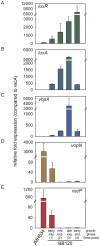
Cells of the wild type (BB120) and the autoinducer-negative mutant JMH634 were grown as described in Fig. 2. Total RNA was isolated at four different time points (marked by the arrows in Fig. 2A), which are characterized by different concentrations/blends of the AIs: 1– early exponential growth phase  =  low concentration of AI-2; 2– mid-exponential growth phase  =  high concentration of AI-2; 3– late exponential growth phase  =  blend of AI-2 and HAI-1; 4– stationary phase  =  blend of AI-2, HAI-1 and CAI-1. Levels of luxR (A), luxA (B), vhpA (C), vopN (D), vscP (D) and recA (as reference) transcripts were determined by qRT-PCR for each time point. Changes in transcript levels (expressed relative to recA) were calculated using the CT method . Since transcript levels of the corresponding genes in mutant JMH634 did not change significantly over time, only one time point (3) is shown. All experiments were performed in triplicate, and error bars indicate standard deviations of the mean.
Figure 7
Figure 7. Phosphorylation activity of LuxQ.
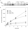
Inverted membrane vesicles prepared from E. coli TKR2000 containing full-length LuxQ were incubated with purified LuxP, purified LuxU and (where indicated) with 10 μM AI-2 (A). The phosphorylation reaction was started by adding 100 μM [γ-32P] ATP at time 0. At the indicated times, the reaction was terminated, and radiolabeled proteins were separated by SDS-PAGE, and visualized by autoradiography. The arrow indicates phosphorylated LuxU. Phosphorylated LuxU was quantified with ImageQuant using [γ-32P] ATP as standard (B). Phosphorylation experiments were also performed in the presence or absence of 10 μM HAI-1 using membrane vesicles containing full-length LuxN and phosphorylated LuxU was quantified accordingly (B).
Figure 8
Figure 8. Effects of various concentrations of HAI-1 and AI-2 on LuxN- and LuxQ- mediated phosphorylation of LuxU.
LuxN- and LuxQ-bearing membrane vesicles, together with purified LuxP and LuxU, were incubated with 100 μM [γ-32P] ATP, and the effects of AI-2 and HAI-1 on the initial rate of LuxU phosphorylation were tested. AI-2 and HAI-1 were added at physiological concentrations (see Fig. 2), indicated in the lower part of the graph (HAI-1 in black, AI-2 in gray). Phosphorylated LuxU was quantitatively analyzed as described in Fig. 7. The degree of inhibition is expressed as the percentage reduction in the initial rate of LuxU phosphorylation measured in the presence of the indicated concentrations/blends of autoinducers relative to that seen in the absence of autoinducers. All experiments were performed in triplicate, and error bars indicate standard deviations of the mean.

References

    1. Fuqua WC, Winans SC, Greenberg EP (1994) Quorum sensing in bacteria: the LuxR-LuxI family of cell density-responsive transcriptional regulators. J Bacteriol 176: 269–275. - PMC - PubMed
    1. Lin B, Wang Z, Malanoski AP, O'Grady EA, Wimpee CF, et al. (2010) Comparative genomic analysis identify the Vibrio harveyi genome sequenced strains BAA-1116 and HY01 as Vibrio campbellii . Environ Microbiol Rep 2: 81–89. - PMC - PubMed
    1. Cano-Gomez A, Høj L, Owens L, Andreakis N (2011) Multilocus sequence analysis provides basis for fast and reliable identification of Vibrio harveyi-related species and reveals previous misidentification of important marine pathogens. Syst Appl Microbiol 34: 561–565. - PubMed
    1. Waters CM, Bassler BL (2005) Quorum sensing: cell-to-cell communication in bacteria. Annu Rev Cell Dev Biol 21: 319–346. - PubMed
    1. Chen X, Schauder S, Potier N, Van DA, Pelczer I, et al. (2002) Structural identification of a bacterial quorum-sensing signal containing boron. Nature 415: 545–549. - PubMed

Publication types

MeSH terms

Substances