. 2013 Feb:40:122-33.
doi: 10.1016/j.jaut.2012.09.003. Epub 2012 Oct 27.

A link between interferon and augmented plasmin generation in exocrine gland damage in Sjögren's syndrome

Affiliations

A link between interferon and augmented plasmin generation in exocrine gland damage in Sjögren's syndrome

Maria Gliozzi et al. J Autoimmun. 2013 Feb.

Abstract

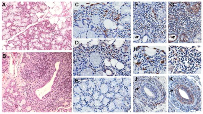
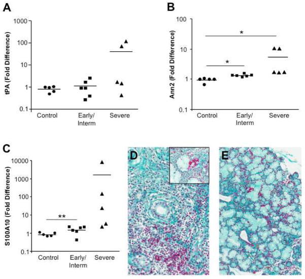
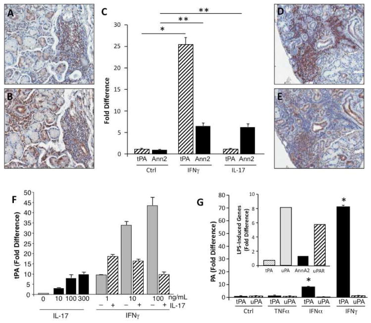
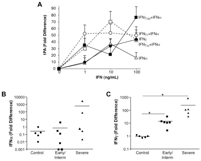
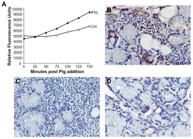
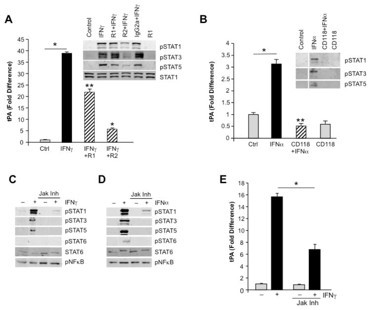
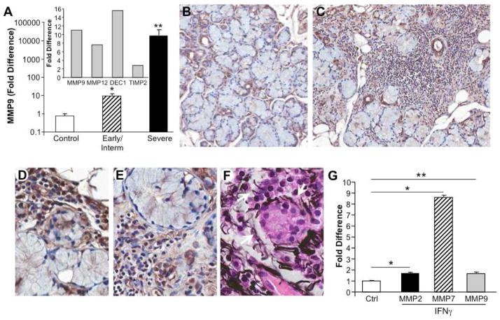
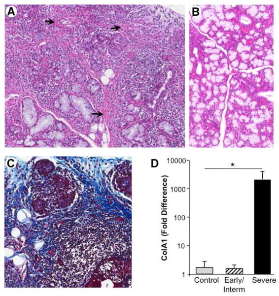
Sjögren's syndrome is an autoimmune disease that targets exocrine glands, but often exhibits systemic manifestations. Infiltration of the salivary and lacrimal glands by lymphoid and myeloid cells orchestrates a perpetuating immune response leading to exocrine gland damage and dysfunction. Th1 and Th17 lymphocyte populations and their products recruit additional lymphocytes, including B cells, but also large numbers of macrophages, which accumulate with disease progression. In addition to cytokines, chemokines, chitinases, and lipid mediators, macrophages contribute to a proteolytic milieu, underlying tissue destruction, inappropriate repair, and compromised glandular functions. Among the proteases enhanced in this local environment are matrix metalloproteases (MMP) and plasmin, generated by plasminogen activation, dependent upon plasminogen activators, such as tissue plasminogen activator (tPA). Not previously associated with salivary gland pathology, our evidence implicates enhanced tPA in the context of inflamed salivary glands revolving around lymphocyte-mediated activation of macrophages. Tracking down the mechanism of macrophage plasmin activation, the cytokines IFNγ and to a lesser extent, IFNα, via Janus kinase (JAK) and signal transducer and activator of transcription (STAT) activation, were found to be pivotal for driving the plasmin cascade of proteolytic events culminating in perpetuation of the inflammation and tissue damage, and suggesting intervention strategies to blunt irreversible tissue destruction.


Figures

Figure 1
Figure 1. Salivary gland infiltration and plasminogen activators
A) Control minor salivary gland illustrating acini, ducts and minimal periductal infiltration. H&E, original magnification 10X. B) Extensive mononuclear cell infiltration and tissue disruption in Sjögren’s syndrome MSG lead to compromised secretory function. Periductal lymphoid infiltrate extends into the acini and throughout parenchyma. Original magnification 10X. C) Immunohistochemical staining for CD68 positive macrophages in representative SS MSG indicates myeloid cells in the mononuclear cell infiltrate. 40X. D) Staining for a tissue component of the plasminogen activation system, tissue plasminogen activator (tPA), demonstrates positive staining in regions of inflammatory cell infiltrate and in some ductal cells. E) Limited evidence of tPA staining in noninflamed MSG tissues. F,G) Salivary gland tissue from SS patient with FS of 7 stained for CD68 (F) and tPA (G) showing staining in infiltrate and ducts. H,I) Higher magnification of CD68 (H) and tPA (I) staining from SS patient with FS = 9.5. J,K) SS (FS=10) MSG tissue stained for CD68 macrophages (J) (arrow denotes CD68+ macrophages appearing to invade duct) which are stained with tPA (K, arrow), but ductal cells are also tPA positive (K).
Figure 2
Figure 2. Enhanced plasmin activation system in inflamed salivary glands
MSG tissues from control subjects (n=5), pSS subjects with early/intermediate disease (n=6) and those with severe SS (n=5) were tested by RT-PCR for tissue plasminogen activator and two cell binding partners. A) RT-PCR for tPA revealed increased plasminogen activator expression in individuals with severe SS. Data (expression normalized to GAPDH) represent fold difference compared to non-SS MSG. B) Increased expression of annexin A2 was modest, but significant in the diseased tissues, and S100A10 was also significantly elevated in early/intermediate and severe disease relative to control (non-SS) tissues (C), *p≤0.01; **p=0.06, Mann-Whitney, two-tailed. D,E) Fraaser-Lendrum staining for fibrin in which fibrin, keratin, and some cytoplasmic granules appear red, erythrocytes appear orange, and matrix is green in MSG from a SS patient (FS=10); original magnification 20X and inset 40X (D) and normal MSG (20X) (E).
Figure 3
Figure 3. Cytokine regulation of components of plasminogen activation
A,B) Representative MSG tissues from patients with SS were stained for CD3 (A) and IFNγ (B) and demonstrate IFNγ+ cells in the inflamed tissues characterized by abundant infiltrating CD3+ cell populations. Original magnification 20X. C) Peripheral blood monocytes were treated with IFNγ (10ng/ml) or IL-17 (100ng/ml) and monitored for altered expression of components of the tPA plasminogen activation system by RT-PCR. IFNγ was an effective inducer of monocyte tPA (*p=0.004 compared to control/unstimulated cells) and IFNγ and IL-17 induced annexin A2 (**p<0.02); n=4. P values calculated using independent Vassar Stats, T test (two-tailed, equal variances). D,E) Representative MSG tissues from patients with SS were stained for CD3 (D) and IL-17 (E) and reveal IL-17+ cells in the inflamed tissues characterized by abundant infiltrating CD3+ cell populations. F) Monocytes were untreated (0), treated with IL-17 (10–300ng/ml), treated with IFNγ (1–100ng/ml) or treated with IFNγ (1–100ng/ml) in the presence of a fixed amount of IL-17 (10ng/ml) and monitored by RT-PCR for tPA expression. G) Comparison of cytokine mediation of tPA and uPA in monocyte-derived macrophages was assessed by RT-PCR. TNFα did not enhance plasminogen activators and IFNγ induced tPA to a greater extent than IFNα (*p=0.001, relative to control, unstimulated macrophages). Inset. LPS (50ng/ml) engagement of TLR triggers components of the uPA system, but not the tPA pathway in macrophages.
Figure 4
Figure 4. IFN enhancement of tPA in vitro and elevated expression in vivo
A) Monocytes were untreated (0), treated with IFNγ only (1–100ng/ml), treated with IFNα only (1–100ng/ml) or treated with IFNγ (1–100ng/ml) in the presence of IFNα (1–100ng/ml) and monitored by RT-PCR for tPA expression. B,C) By RT-PCR, MSG tissues from non-SS MSG, SS patients with early/intermediate disease and severe disease exhibited variable IFNα levels with progressive disease (B), whereas significant increases in expression of IFNγ were evident in early/intermediate disease and in severe disease (*p≤0.01) as compared to control non-SS tissues (C). P values calculated using Vassar Stats, Mann Whitney (two-tailed).
Figure 5
Figure 5. Enhanced plasminogen activation by IFNγ in vitro and increased plasmin in vivo
A) Monocytes were cultured in the presence or absence of IFNγ and tested for their ability to activate plasminogen resulting in plasmin formation as detected by proteolytic activity and cleavage of a colorimetric substrate (relative fluorescence units) over time, as indicated. B) Representative MSG tissues stained with an antibody to plasmin(ogen) reveal cell associated and intercellular positive staining within the inflammatory infiltrate, endothelial and ductal cells, relative to an isotype control antibody (C) and compared to control non-SS gland tissues (D). Original magnification 40X.
Figure 6
Figure 6. Signaling pathways in macrophage plasminogen activation
A) Macrophage cultures were stimulated or not with IFNγ in the presence or absence of antibodies to the IFNγ receptor (R1, R2) and tPA expression by RT-PCR and inset) p-STAT signaling monitored by Western blot. IFNγ significantly enhanced tPA relative to control, unstimulated macrophages (A, *p<0.001). As evident, phosphorylations of STAT1 and particularly, STAT3 and STAT5 were inhibited in the presence of antibody to R2 (inset), which resulted in significantly blunted expression of tPA relative to IFNγ-only treated macrophages. *p<0.001; **p<0.01. B. Macrophage cultures were stimulated or not with IFNα in the presence or absence of antibodies to IFNAR (CD118) and tPA levels monitored by RT-PCR (B) and p-STAT signaling monitored by Western blot (inset). Phosphorylations of STAT1, STAT3 and STAT5 were all inhibited in the presence of the antibody, which resulted in significant blockage of tPA expression relative to IFNα. *p<0.01. C,D) Using a Jak1/2 inhibitor, both IFNα and IFNγ signaling pathways were interrupted with near loss of STAT phosphorylation as determined by Western blot using STAT6 protein as loading control. No inhibition was seen on constitutive pNFκB. E) In the presence of the Jak inhibitor, IFNγ-induced tPA expression was significantly inhibited. *p=0.01 comparing IFNγ only with IFNγ + Jak Inh.
Figure 7
Figure 7. MMP expression in MSG
A) By microarray, SS salivary glands with severe lesions exhibited enhanced MMP9, MMP12 and ADAMDEC1 expression relative to gland tissues from subjects without SS and minimally increased TIMP2 (inset). By RT-PCR, MSG tissues from additional populations (n=5–6/group) were analyzed for expression of MMP9. By comparison to non-SS MSG, those tissues with early/intermediate disease exhibited a significant elevation in MMP9 expression (*p=0.004), with a more dramatic elevation in the severely inflamed and damaged MSG (**p=0.006). B) Immunohistochemical staining for MMP9 in control MSG revealed staining in ductal cells and/or in the periductal regions. Original magnification 20X. C,D,E) Compared to control MSG, tissues obtained from patients with severe SS exhibited extensive staining for MMP in the infiltrate and around acini (C, original magnification 20X), consonant with basal lamina damage, detachment of acinar cells, loss of nuclear polarity and disrupted structural integrity (D, E). Original magnification 40X. F) Immunohistochemistry using Jone’s PAMS stain for basement membrane identifies areas of fragmented basement membrane, particularly evident around acinar structures (white arrows). Original magnification 40X. G) Monocytes were cultured in the presence or absence of IFNγ (10ng/ml) for 4hr and MMP2, MMP7 and MMP9 expression monitored by RT-PCR. * p<0.01, **p=0.05 compared to control unstimulated macrophages (no IFNγ).
Figure 8
Figure 8. Loss of salivary gland integrity and matrix accumulation
A) In MSG from patients with advanced disease, the extensive lymphoid infiltrate with germinal centers leads to interstitial fibrosis (arrows) and acinar atrophy, as ECM and adipose cells impinge on structure and functionality, compared to noninflamed MSG (B) H&E, original magnification 10X. C) Trichrome stain delineating interstitial fibrosis, along with acinar loss and mononuclear infiltration. Original magnification 20X. D) By RT-PCR, a representative collagen molecule, Col1A1, expression was not increased in early/intermediate disease MSG, but was significantly enhanced in the MSG of subjects with chronic/severe SS relative to MSG of individuals without SS; p<0.01.

References

    1. Voulgarelis M, Dafni UG, Isenberg DA, Moutsopoulos HM. Malignant lymphoma in primary Sjogren’s syndrome: a multicenter, retrospective, clinical study by the European Concerted Action on Sjogren’s Syndrome. Arthritis Rheum. 1999;42:1765–72. - PubMed
    1. Ice JA, Li H, Adrianto I, Lin PC, Kelly JA, Montgomery CG, et al. Genetics of Sjögren’s syndrome in the genome-wide association era. J Autoimmun. 2012;39:57–63. - PMC - PubMed
    1. Katsifis GE, Moutsopoulos NM, Wahl SM. T lymphocytes in Sjogren’s syndrome: contributors to and regulators of pathophysiology. Clin Rev Allergy Immunol. 2007;32:252–64. - PubMed
    1. Manoussakis MN, Boiu S, Korkolopoulou P, Kapsogeorgou EK, Kavantzas N, Ziakas P, et al. Rates of infiltration by macrophages and dendritic cells and expression of interleukin-18 and interleukin-12 in the chronic inflammatory lesions of Sjogren’s syndrome: correlation with certain features of immune hyperactivity and factors associated with high risk of lymphoma development. Arthritis Rheum. 2007;56:3977–88. - PubMed
    1. Greenwell-Wild T, Moutsopoulos NM, Gliozzi M, Kapsogeorgou E, Rangel Z, Munson PJ, et al. Chitinases in the salivary glands and circulation of patients with Sjogren’s syndrome: Macrophage harbingers of disease severity. Arthritis Rheum. 2011;63:3103–15. - PMC - PubMed

Publication types

MeSH terms