. 2013 Jan 15;373(2):383-93.
doi: 10.1016/j.ydbio.2012.10.023. Epub 2012 Oct 27.

The UPD3 cytokine couples environmental challenge and intestinal stem cell division through modulation of JAK/STAT signaling in the stem cell microenvironment

Affiliations

The UPD3 cytokine couples environmental challenge and intestinal stem cell division through modulation of JAK/STAT signaling in the stem cell microenvironment

Feng Zhou et al. Dev Biol. .

Abstract

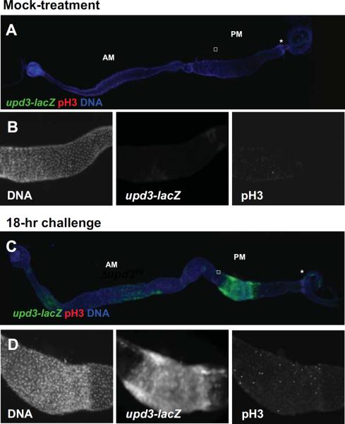
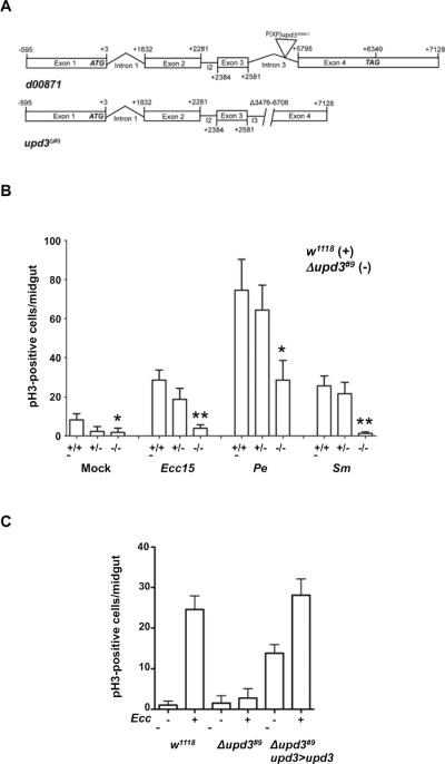
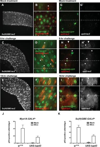
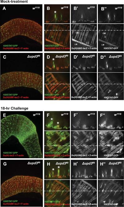
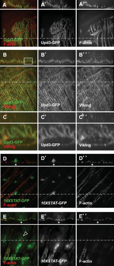
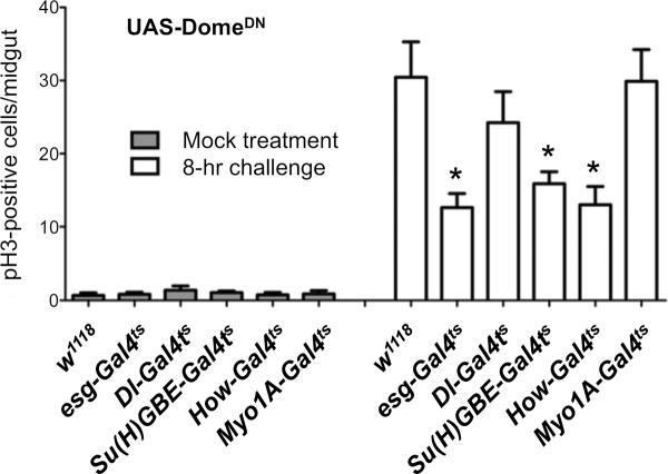
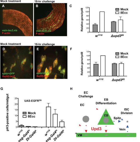
In Drosophila, the replacement of spent enterocytes (ECs) relies on division of intestinal stem cells (ISCs) and differentiation of their progeny, the enteroblasts (EBs). Recent studies have revealed a role for JAK/STAT signaling in the modulation of the rate of ISC division in response to environmental challenge. Here, we demonstrate the critical role of the UPD3 cytokine in the JAK/STAT-dependent response to enteric infection. We show that upd3 expression is activated in ECs and in EBs that massively differentiate in response to challenge. We show that the UPD3 cytokine, which is secreted basally and accumulates at the basement membrane, is required for stimulation of JAK/STAT signaling in EBs and visceral muscles (VMs). We further show that stimulation of ISC division requires active JAK/STAT signaling in EBs and VMs, but apparently not in ISCs. Our results suggest that EBs and VMs modulate the rate of the EGFR-dependent ISC division through upd3-dependent production of the EGF ligands Spitz and Vein, respectively. This study therefore supports the notion that the production of the UPD3 cytokine in stem cell progeny (ECs and EBs) stimulates intestinal stem cell division through modulation of JAK/STAT signaling in the stem cell microenvironment (EBs and VMs).


Figures

Fig. 1
Fig. 1. upd3 is expressed in the posterior midgut in response to challenge
(A–D) Fluorescence images showing upd3-lacZ expression and pH3-positive nuclei in response to mock-treatment (A and B) and 18-hour Ecc challenge (C and D). Whole intestine are shown in (A) and (C). (B) and (D) correspond to magnification of the squared regions displayed in (A) and (C), respectively. AM: anterior midgut; PM: posterior midgut; asterisk: hindgut proliferating zone.
Fig. 2
Fig. 2. The Upd3 cytokine is required for stimulation of ISC division in response to challenge
(A) Structural organization of the upd3 locus in the parental strain (d00871) and in the upd3 deletion mutant (Δupd3#9). (B) pH3-positive nuclei were scored in wild-type (w1118), heterozygote (Δupd3#9/w1118), and upd3 mutant (Δupd3#9) animals infected for 18 hours with Erwinia carotovora caratovora 15 (Ecc15) (OD600=50), Pseudomonas entomophila (Pe), or Serratia marcescens (Sm). Averages of three representative experiments (n = 12 guts/experiment) with standard deviation are shown. P values were calculated by two-tailed, unpaired t-tests. For Δupd3#9 vs. w1118: mock, P = 0.0437 (*); Ecc15 infection, P = 0.0015 (**); Pe infection, P = 0.0132 (*); Sm infection, P = 0.0010 (**). For infection vs. mock, w1118: Ecc15, P =0.0044 (**); Pe, P = 0.0021 (**); Sm, P = 0.0066 (**). For infection vs. mock, Δupd3#9: Ecc15, P =0.2698 (ns); Pe, P = 0.0111 (*); Sm, P = 0.7408 (ns). (C) Genetic rescue of the upd3 mutant. pH3-positive nuclei were scored in wild-type (w1118), upd3 mutant (Δupd3#9) or rescued upd3 mutant animals (Δupd3#9;upd3>upd3: ectopic expression of UAS-Upd3 with upd3-GAL4, tub-Gal80ts). P values were calculated by two-tailed, unpaired t-tests. For mock infection, Δupd3#9 vs. w1118, P =0.2995 (ns) and wild-type vs. rescue, P<0.0001 (***). For Ecc15 infection, Δupd3#9 vs. w1118, P<0.0001 (***) and upd3>upd3 vs. w1118, P =0.38 (ns).
Fig. 3
Fig. 3. upd3 is expressed and required in differentiated ECs and in differentiating EBs
(A, C and E) Confocal microscopy images of posterior midgut (10× objective, XY section). (B, D, F, G, H and I) Confocal microscopy images of posterior midgut (60× objective). Bottom panels show XY section. Dotted lines indicate Y coordinates for XZ reconstruction (top panels). (A–F) Expression of the EC-specific marker Myo1A and EB-specific marker Su(H)-lacZ in mock-treated animal (A and B), animals infected for 8 hours with Ecc15 (C and D) and animals infected for 18 hours with Ecc15 (E and F). (G–I) Expression of the EB-specific marker Su(H)>GFP and the upd3-lacZ reporter in mock-treated animal (G), animals infected for 8 hours with Ecc15 (H) and animals infected for 18 hours with Ecc15 (I). (J and K) pH3-positive nuclei were scored in Myo1A-Gal4ts/+ (w1118) or Myo1A-Gal4ts/UAS-iupd3 (UAS-iupd3) (J) and Su(H)-GAL4ts/+ (w1118) or Su(H)-Gal4ts/UAS-iupd3 (UAS-iupd3) (K) animals infected with Ecc15 for 8 hours. Averages of three representative experiments (n = 12 guts/experiments) with s.d. are shown. (J) UAS-iupd3 vs. w1118, P = 0.0462 (*). (K) UAS-iupd3 vs. w1118, P = 0.0164 (*).
Fig. 4
Fig. 4. Upd3 is required for activation of JAK/STAT signaling in EBs and visceral muscles in response to challenge
(A–H) Low-magnification (left panels) and high-magnification (right panels) confocal microscopy images of 10XSTAT-GFP and Su(H)GBE-lacZ expression in the posterior midgut. Bottom right panels show XY sections. Dotted lines indicate Y coordinates for XZ reconstruction (top right panels). Images corresponding to mock-treated (A and B) and challenged (E and F) wild-type (w1118) and mock-treated (C and D) and challenged (G and H) upd3 mutant (Δupd3#9) animals.
Fig. 5
Fig. 5. The Upd3 cytokine produced in EB locates to the extracellular matrix and activates JAK/STAT signaling in EB and in visceral muscles
(A) The Upd3-GFP fusion decorates muscles as visualized by F-actin staining. (B) The Upd3-GFP fusion protein co-localizes with the extracellular matrix marker Viking. (C) Magnification of the squared area shown in (B). (D) The 10X-STAT-GFP reporter is expressed in EBs and display basal expression level in visceral muscles. (E) Over-expression of the UAS-upd3 transgene using the Su(H)-Gal4ts driver leads to 10X-STAT-GFP expression in visceral muscles (asteriks) and differentiation of EBs (arrowhead pointing to the nucleus of a differentiating EBs).
Fig. 6
Fig. 6. JAK/STAT signaling is required in enteroblasts and visceral muscles for ISC division in response to challenge
Expression of a dominant negative version of the Domeless receptor (UAS-domeΔCYT) in ISCs and EBs (esg-GAL4), ISCs alone (Dl-GAL4), EBs alone (Su(H)GBE-GAL4), visceral muscles (how-GAL4), or enterocytes (Myo1A-GAL4). Animals were mock-treated (Mock) or infected with Ecc15 for 8 hours (8Ecc). Averages of six independent experiments (n = 12 guts/experiments) with s.e.m. are shown. P values were calculated by two-tailed, unpaired t-tests. Compared to control, esg-GAL4, P = 0.012 (*); Dl-GAL4, P = 0.36 (ns); Su(H)GBE-GAL4, P = 0.017 (*), how-GAL4, P = 0.015 (*); Myo1A-GAL4, P = 0.93 (ns).
Fig. 7
Fig. 7. Role of Upd3 signaling in EGFR-dependent ISC division
(A–B) Vein is weakly expressed in visceral muscles as visualized by F-actin staining in mock-treated animals and highly expressed in animals infected for 18 hours with Ecc15. (C) Quantification of vein expression by RT-PCR in mock-treated animals and in animals infected with Ecc15 for 8 hours (8Ecc). For w1118, 8Ecc vs. Mock, P = 0.0006 (***). For Δupd3#9, 8Ecc vs. Mock, P = 0.5585 (ns). (D and E) Spitz is expressed in EBs as visualized with the Su(H)GBE-lacZ marker in mock-treated animals and in differentiated EBs in animals infected with Ecc15 for 18 hours. (F) Quantification of spitz expression by RT-PCR in mock-treated animals and in animals infected with Ecc15 for 8 hours (8Ecc). For w1118, 8Ecc vs. Mock, P = 0.0101 (*). For Δupd3#9, 8Ecc vs. Mock, P = 0.8212 (ns). (G) EGF signaling was conditionally (using tub-GAL80ts) inhibited in ISCs and EBs (esg-GAL4), or ISCs alone (Dl-GAL4) by expressing a dominant-negative form of the EGF receptor (UAS-EGFRDN). For Ecc15 treatment (8Ecc), escg-GAL4 vs. w1118, P = 0.0414 (*) and Dl-GAL4 vs. w1118, P = 0.0012 (**). (H) Model of intestinal homeostasis. Erwinia infection activates upd3 expression in mature enterocytes (EC) and in differentiating enteroblasts (EB) (Upd3 production and producing cells are depicted in red). The basal secretion of the Upd3 cytokine activates JAK/STAT signaling in EBs and in visceral muscles (VM) (JAK/STAT signaling depicted in green). Activation of JAK/STAT signaling in EBs and VMs leads to EGF ligand production that stimulates the EGFR-dependent division of ISCs (EGF signaling depicted in blue).

References

    1. Amcheslavsky A, Jiang J, Ip YT. Tissue damage-induced intestinal stem cell division in Drosophila. Cell Stem Cell. 2009;4:49–61. - PMC - PubMed
    1. Apidianakis Y, Pitsouli C, Perrimon N, Rahme L. Synergy between bacterial infection and genetic predisposition in intestinal dysplasia. Proc Natl Acad Sci U S A. 2009;106:20883–20888. - PMC - PubMed
    1. Bach EA, Ekas LA, Ayala-Camargo A, Flaherty MS, Lee H, Perrimon N, Baeg GH. GFP reporters detect the activation of the Drosophila JAK/STAT pathway in vivo. Gene Expr Patterns. 2007;7:323–331. - PubMed
    1. Basset A, Khush RS, Braun A, Gardan L, Boccard F, Hoffmann JA, Lemaitre B. The phytopathogenic bacteria Erwinia carotovora infects Drosophila and activates an immune response. Proc Natl Acad Sci U S A. 2000;97:3376–3381. - PMC - PubMed
    1. Becker C, Fantini MC, Schramm C, Lehr HA, Wirtz S, Nikolaev A, Burg J, Strand S, Kiesslich R, Huber S, Ito H, Nishimoto N, Yoshizaki K, Kishimoto T, Galle PR, Blessing M, Rose-John S, Neurath MF. TGF-beta suppresses tumor progression in colon cancer by inhibition of IL-6 trans-signaling. Immunity. 2004;21:491–501. - PubMed

Publication types

MeSH terms