. 2012 Dec 18;44(24):1188-200.
doi: 10.1152/physiolgenomics.00097.2012. Epub 2012 Oct 30.

Glucocorticoid treatment of astrocytes results in temporally dynamic transcriptome regulation and astrocyte-enriched mRNA changes in vitro

Affiliations

Glucocorticoid treatment of astrocytes results in temporally dynamic transcriptome regulation and astrocyte-enriched mRNA changes in vitro

Bradley S Carter et al. Physiol Genomics. .

Abstract

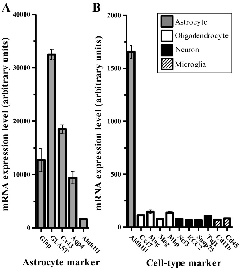
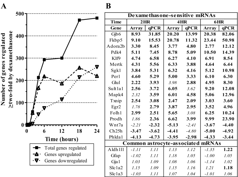
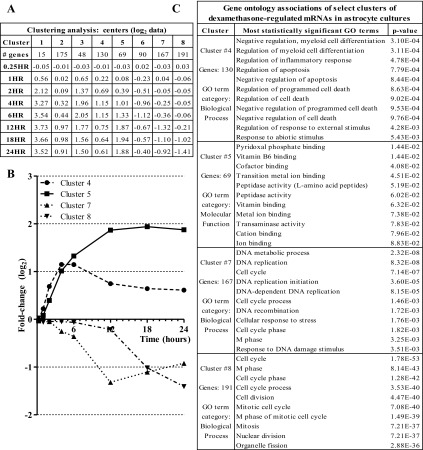
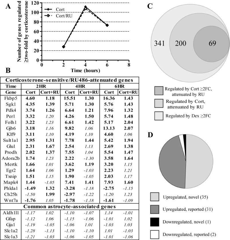
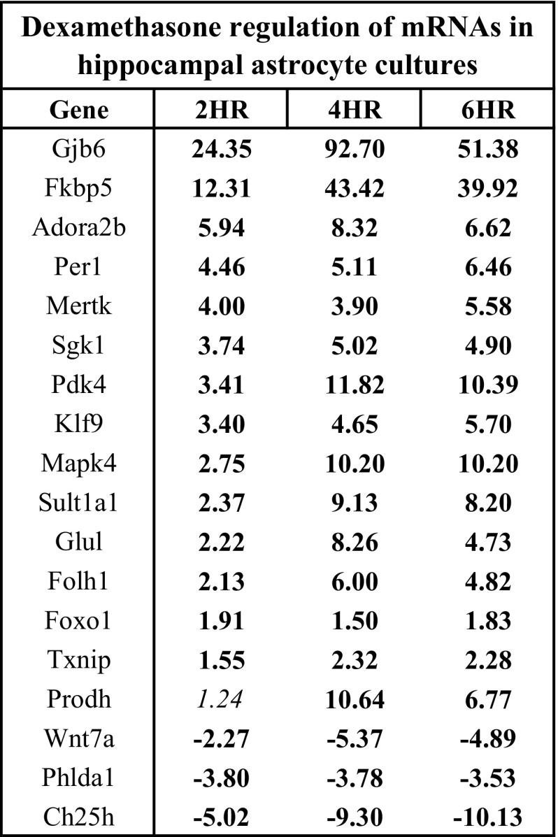
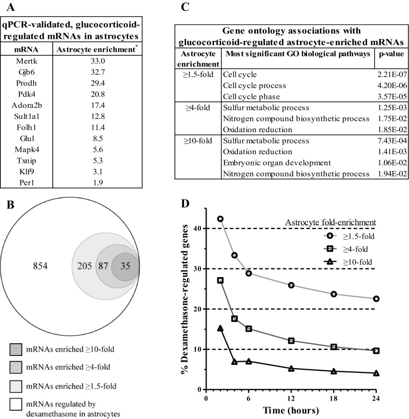
While general effects of glucocorticoids are well established, the specific cellular mechanisms by which these hormones exert tissue-dependent effects continue to be elaborated. Diseases that demonstrate altered glucocorticoid signaling have been associated with alterations in astrocytes, yet relatively little is known about the effects of glucocorticoids upon this cell type. We have analyzed mRNA expression patterns following glucocorticoid treatment of mouse primary astrocyte cultures. Microarray analysis of cortical astrocyte cultures treated with dexamethasone over an eight-point, 24 h time course identified 854 unique genes with ≥twofold change in mRNA expression at one or more time points. Clustering analysis associated subsets of these mRNA expression changes with gene ontology categories known to be impacted by glucocorticoids. Numerous mRNAs regulated by dexamethasone were also regulated by the natural ligand corticosterone; all of the mRNAs regulated ≥twofold by corticosterone were substantially attenuated by cotreatment with the glucocorticoid receptor antagonist RU486. Of the mRNAs demonstrating ≥twofold expression change in response to both glucocorticoids, 33 mRNAs were previously associated with glucocorticoid regulation, and 36 mRNAs were novel glucocorticoid targets. All genes tested by qPCR for glucocorticoid regulation in cortical astrocyte cultures were also regulated by glucocorticoids in hippocampal astrocyte cultures (18/18). Interestingly, a portion of glucocorticoid-regulated genes were astrocyte enriched; the percentage of astrocyte-enriched genes per total number of regulated genes was highest for the early time points and steadily decreased over the time course. These findings suggest that astrocytes in vitro may initially deploy cell type-specific patterns of mRNA regulatory responses to glucocorticoids and subsequently activate additional cell type-independent responses.


Figures

Fig. 1.
Fig. 1.
Cell culture system specifically expresses astrocyte mRNA cell markers. A: plot of average mRNA expression for subset of astrocyte cell markers among sample groups from 8 time points in Dexamethasone (Dex) time course. B: plot of average mRNA expression for subset of cell markers associated with additional brain cell types (neurons, oligodendrocytes, microglia). Bars for each mRNA are colored according to the cell type association. Error bars display SE.
Fig. 2.
Fig. 2.
Dex dynamically regulates specific mRNAs in astrocytes in vitro over 24 h time course. A: plot of number of genes that show >2-fold regulation by dexamethasone over time (P < 0.05). B: comparison of linear fold-change mRNA expression values of microarray data with qPCR data for select mRNAs regulated by Dex treatment (vs. vehicle). Data ordered based on magnitude of mRNA expression change in 2 h qPCR data. HR, hours; boldface = P < 0.05; italics = P > 0.05.
Fig. 3.
Fig. 3.
Clustering analysis of Dex gene expression time course data yields distinct sets of genes that contain similar gene ontology (GO) categories. A: table of number of genes per cluster and cluster centers (average log2 value) over the time course. B: graph of 4 GO term-associated cluster centers from k-means cluster analysis. C: table of graphed clusters and their corresponding most significant GO terms.
Fig. 4.
Fig. 4.
mRNAs regulated by synthetic glucocorticoid in primary astrocyte cultures are also regulated by natural ligand and the regulation is attenuated by GR antagonist. A: plot of number of genes that show >2-fold mRNA expression change due to corticosterone (Cort) that is attenuated by RU486 cotreatment (Cort/RU) (P < 0.05). B: table of qPCR validation data on select Cort-sensitive mRNAs. Genes ordered by magnitude of fold-change due to 2 h Cort exposure. C: proportional Venn diagram of overlap of genes regulated >2-fold by Dex at 2, 4, or 6 h vs. genes regulated by Cort (P < 0.05) and attenuated by RU486 (P > 0.05) at those same time points. D: graph characterizing genes regulated by both Dex and Cort ≥2-fold and also attenuated by RU486 in terms of previous literature reporting and direction of regulation. Regulated by Cort = P < 0.05, attenuated by RU = P > 0.05. FC, fold-change; boldface = P < 0.05, italics = P > 0.05.
Fig. 5.
Fig. 5.
A portion of mRNAs regulated by glucocorticoids in cortical astrocyte cultures is also regulated by glucocorticoids in hippocampal astrocyte cultures. qPCR data of select mRNAs regulated by Dex treatment in cortical astrocyte cultures that are also hippocampal astrocyte cultures. Genes ordered based on 2 h FC values. Boldface = P < 0.05, italics = P > 0.05.
Fig. 6.
Fig. 6.
Certain mRNAs regulated by Dex are astrocyte enriched. A: table of genes found to be regulated ≥2-fold by Dex and Cort in both microarray and qPCR experiments and their reported enrichment in astrocytes relative to other neural cell types. Genes arranged by magnitude of enrichment. *Cahoy et. al., (8). B: proportional Venn diagram of overlap of total number of unique Dex-regulated mRNAs with 3 subsets of astrocyte-enriched mRNAs. C: table containing most statistically significant GO category associated with 3 astrocyte-enriched mRNA subsets of Dex-regulated mRNAs in B. D: plot of percentage of astrocyte-enriched mRNAs as a function of total mRNAs regulated ≥2-fold by Dex over time.

References

    1. Abbott NJ, Rönnbäck L, Hansson E. Astrocyte-endothelial interactions at the blood-brain barrier. Nat Rev Neurosci 7: 41–53, 2006. - PubMed
    1. Barley K, Dracheva S, Byne W. Subcortical oligodendrocyte-and astrocyte-associated gene expression in subjects with schizophrenia, major depression and bipolar disorder. Schizophrenia Res 112: 54–64, 2009. - PubMed
    1. Baulieu E. The steroid hormone antagonist RU486. Mechanism at the cellular level and clinical applications. Endocrinol Metab Clin No Am 20: 873, 1991. - PubMed
    1. Bernard R, Kerman IA, Thompson RC, Jones EG, Bunney WE, Barchas JD, Schatzberg AF, Myers RM, Akil H, Watson SJ. Altered expression of glutamate signaling, growth factor, and glia genes in the locus coeruleus of patients with major depression. Mol Psychiatry 16: 634–646, 2010. - PMC - PubMed
    1. Brown AM, Ransom BR. Astrocyte glycogen and brain energy metabolism. Glia 55: 1263–1271, 2007. - PubMed

Publication types

MeSH terms