

Evidence for natural recombination between mink enteritis virus and canine parvovirus
- PMID: 23110843
- PMCID: PMC3495801
- DOI: 10.1186/1743-422X-9-252
Evidence for natural recombination between mink enteritis virus and canine parvovirus
Abstract
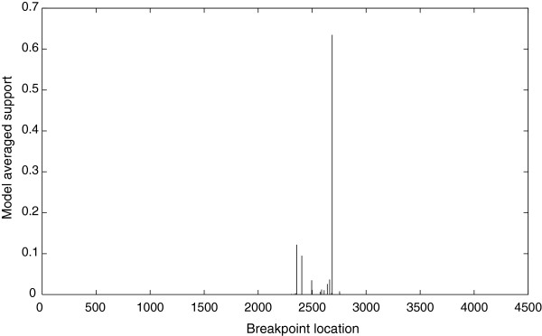
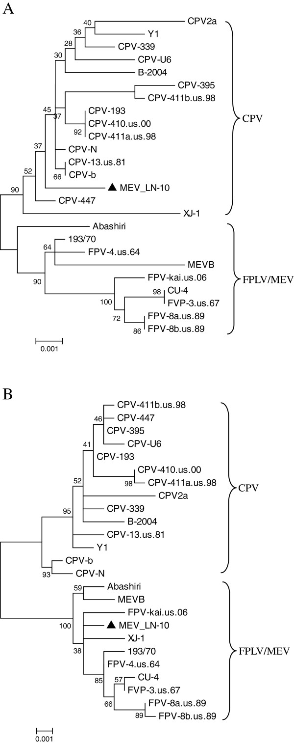
A virus was isolated from mink showing clinical and pathological signs of enteritis in China. This virus, designated MEV/LN-10, was identified as mink enteritis virus (MEV) based on its cytopathic effect in the feline F81 cell line, the hemagglutination (HA) and hemagglutination inhibition (HI) assay, electron microscopy (EM) and animal infection experiments. The complete viral genome was cloned and sequenced. Phylogenetic and recombination analyses on the complete MEV/LN-10 genome showed evidence of recombination between MEV and canine parvovirus (CPV). The genome was composed of the NS1 gene originating from CPV while the VP1 gene was of MEV origin. This is the first demonstration of recombination between a CPV and MEV in nature. Our findings not only provide valuable evidence indicating that recombination is an important genetic mechanism contributing to the variation and evolution of MEV, but also that heterogeneous recombination can occur in the feline parvovirus subspecies.
Figures
References
-
- Schofield FW. Virus enteritis in mink. N Am Vet. 1949;30:651–654.
-
- Steinel A, Parrish CR, Bloom ME, Truyen U. Parvovirus infections in wild carnivores. J Wildl Dis. 2001;37:594–607. - PubMed
Publication types
MeSH terms
Substances
Associated data
- Actions
LinkOut - more resources
Full Text Sources
