Review
. 2013 Feb;199(2):89-98.
doi: 10.1007/s00359-012-0769-3. Epub 2012 Oct 31.

The magnetite-based receptors in the beak of birds and their role in avian navigation

Affiliations
Review

The magnetite-based receptors in the beak of birds and their role in avian navigation

R Wiltschko et al. J Comp Physiol A Neuroethol Sens Neural Behav Physiol. 2013 Feb.

Abstract

Iron-rich structures have been described in the beak of homing pigeons, chickens and several species of migratory birds and interpreted as magnetoreceptors. Here, we will briefly review findings associated with these receptors that throw light on their nature, their function and their role in avian navigation. Electrophysiological recordings from the ophthalmic nerve, behavioral studies and a ZENK-study indicate that the trigeminal system, the nerves innervating the beak, mediate information on magnetic changes, with the electrophysiological study suggesting that these are changes in intensity. Behavioral studies support the involvement of magnetite and the trigeminal system in magnetoreception, but clearly show that the inclination compass normally used by birds represents a separate system. However, if this compass is disrupted by certain light conditions, migrating birds show 'fixed direction' responses to the magnetic field, which originate in the receptors in the beak. Together, these findings point out that there are magnetite-based magnetoreceptors located in the upper beak close to the skin. Their natural function appears to be recording magnetic intensity and thus providing one component of the multi-factorial 'navigational map' of birds.


Figures

Fig. 1
Fig. 1
Electrophysiological recordings from the trigeminal ganglion of the bobolink. Left spontaneous activity and responses to various changes in the intensity of the vertical component of the magnetic field. The lines indicate the 50 ms of stimulation (from Semm and Beason 1990). Right recorded activity changes as a logarithmic function of the increased intensity (from Beason and Semm 1991)
Fig. 2
Fig. 2
Orientation of migrants after a short, strong magnetic pulse. Upper diagrams responses of Australian Silvereyes showing that adult (ad.) birds respond with a marked deflection to the east, whereas juveniles (juv.) are unaffected. Black open symbols: behavior before pulsing; solid red symbols: behavior on the day of pulsing and the following day (data from Munro et al. 1997). Lower diagrams responses of bobolinks to pulses applied in two different directions. The headings are presented in relation to the orientation before the pulse (C) projected upward (data from Beason et al. 1995). The arrows represent the mean vector in relation to the radius of the circle = 1; the two inner circles are the 5 % (dashed) and 1 % significance border of the Rayleigh test
Fig. 3
Fig. 3
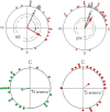
The response to the pulse can be abolished by preventing the information from the magnetite-based receptors reaching the brain—in that case, the birds head in their migratory direction with their magnetic compass, indicating that the pulse does not affect the compass. Upper diagrams response of Bobolinks to a pulse ‘south anterior (P) and when the ophthalmic branch of the trigeminal nerve was anesthetized with a local anesthetic (data from Beason and Semm 1996). Lower diagrams orientation of Australian Silvereyes before pulsing (C, black symbols) and after being subjected to the pulse without further treatment (P, red symbols) and with the upper beak locally anesthetized by a local anesthetic, externally applied (P Xy, blue symbols) (data from W. Wiltschko et al. 2009). Symbols as in Fig. 2
Fig. 4
Fig. 4
‘Fixed direction’ responses in Robins, observed under certain light regimes, can be disrupted by local anesthesia of the upper beak. D, orientation in total darkness; GY, orientation under a combination of monochromatic green and yellow light. Black open symbols: untreated controls; blue solid symbols: birds with the upper beak locally anesthetized with an externally applied local anesthetic (data from Stapput et al. ; R. Wiltschko et al. 2012). Symbols as in Fig. 2
Fig. 5
Fig. 5
Vanishing intervals of untreated pigeons (Cont., controls) and pigeons with the upper beak anesthetized by an externally applied local anesthetic (Exp., experimentals) within an strong, irregular anomaly and outside in magnetically quiet terrain. The columns indicate mean and standard deviations. Asterisks indicate significant differences: *p < 0.05; ***p < 0.001 (data from R. Wiltschko et al. 2010a, b)

References

    1. Beason RC, Brennan WJ. Natural and induced magnetization in the Bobolink Dolichonyx oryzivorus (Aves: Icteridae) J Exp Biol. 1986;125:49–56.
    1. Beason RC, Nichols JE. Magnetic orientation and magnetically sensitive material in a transequatorial migratory bird. Nature. 1984;309:151–153. doi: 10.1038/309151a0. - DOI
    1. Beason RC, Semm P. Magnetic responses of the trigeminal nerve system of the Bobolink (Dolichonyx oryzivorus) Neurosci Lett. 1987;80:229–234. doi: 10.1016/0304-3940(87)90659-8. - DOI - PubMed
    1. Beason RC, Semm P. Two different magnetic systems in avian orientation. In: Bell BD, Cossee RO, Flux JEC, Heather BD, Hitchmough RA, Robertson CJR, Williams MJ, editors. Acta XX Congr Int Ornithol. Wellington: New Zealand Ornithol Congr Trust Board; 1991. pp. 1813–1819.
    1. Beason RC, Semm P. Does the avian ophthalmic nerve carry magnetic information? J Exp Biol. 1996;199:1241–1244. - PubMed

MeSH terms

Substances

LinkOut - more resources