Clinical Trial
. 2012 Nov 13;109(46):18885-90.
doi: 10.1073/pnas.1205785109. Epub 2012 Oct 29.

Immunoglobulin-like transcript receptors on human dermal CD14+ dendritic cells act as a CD8-antagonist to control cytotoxic T cell priming

Affiliations
Clinical Trial

Immunoglobulin-like transcript receptors on human dermal CD14+ dendritic cells act as a CD8-antagonist to control cytotoxic T cell priming

Jacques Banchereau et al. Proc Natl Acad Sci U S A. .

Abstract

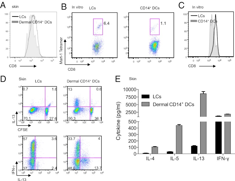
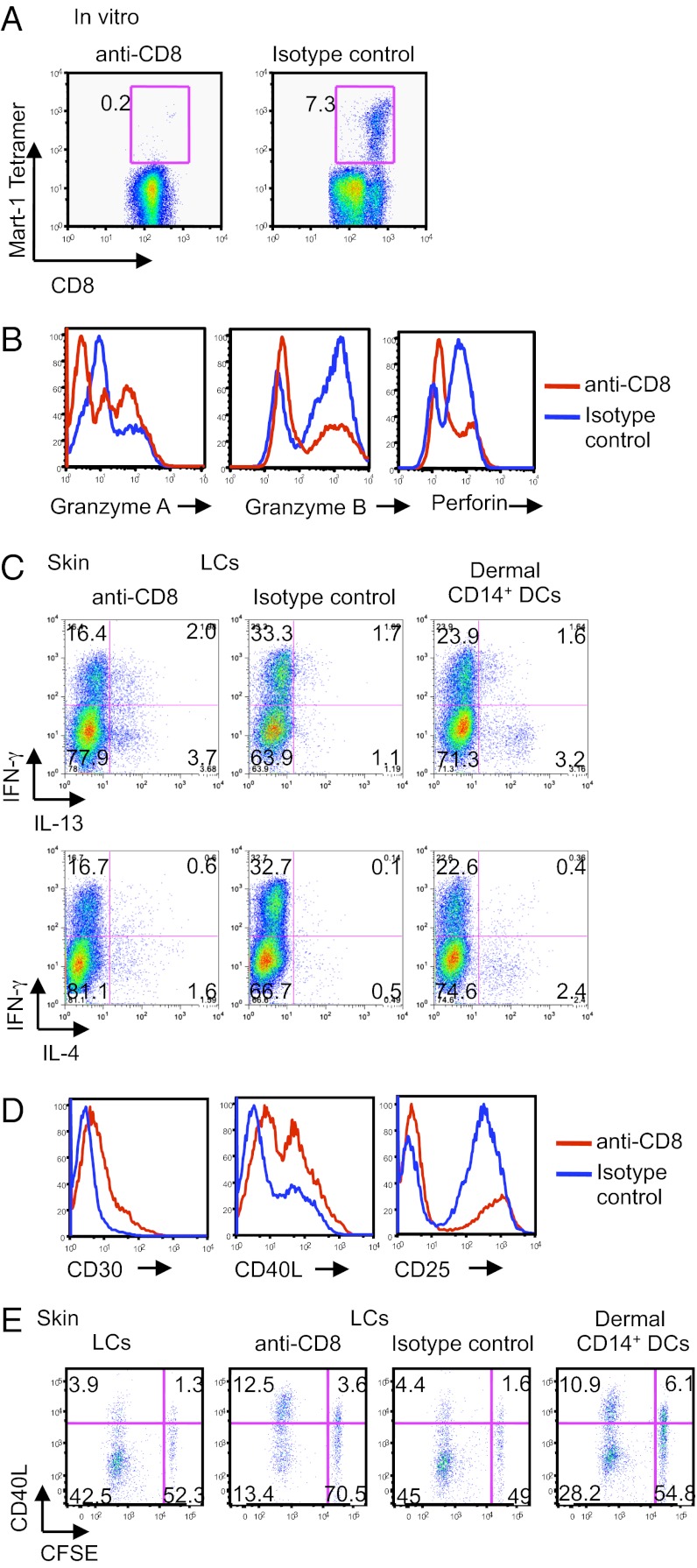
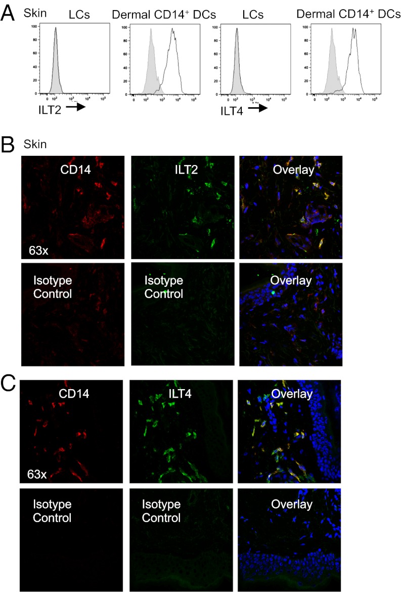
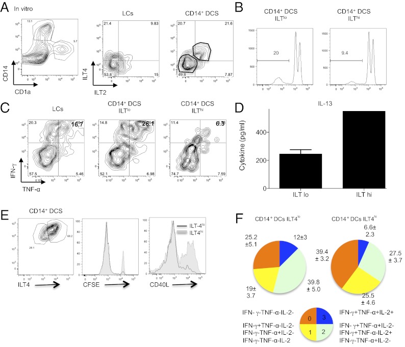
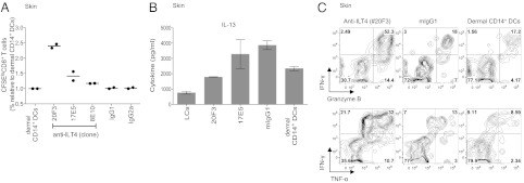
Human Langerhans cells (LCs) are highly efficient at priming cytolytic CD8(+) T cells compared with dermal CD14(+) dendritic cells (DCs). Here we show that dermal CD14(+) DCs instead prime a fraction of naïve CD8(+) T cells into cells sharing the properties of type 2 cytokine-secreting CD8(+) T cells (TC2). Differential expression of the CD8-antagonist receptors on dermal CD14(+) DCs, the Ig-like transcript (ILT) inhibitory receptors, explains the difference between the two types of DCs. Inhibition of CD8 function on LCs inhibited cytotoxic T lymphocytes (CTLs) and enhanced TC2 generation. In addition, blocking ILT2 or ILT4 on dermal CD14(+) DCs enhanced the generation of CTLs and inhibited TC2 cytokine production. Lastly, addition of soluble ILT2 and ILT4 receptors inhibited CTL priming by LCs. Thus, ILT receptor expression explains the polarization of CD8(+) T-cell responses by LCs vs. dermal CD14(+) DCs.


Conflict of interest statement

Conflict of interest statement: J.B., K.A.P., G.Z., and E.K. are co-inventors on a patent filing related to this work.

Figures

Fig. 1.
Fig. 1.
Dermal CD14+ DCs prime CD8low TC2 cells. (A) Naïve CD8+ T cells were primed for 7 d by CD40L-activated skin isolated DC subsets: LCs (black) and dermal CD14+ DCs (gray). CFSElo cells were analyzed by flow cytometry for the expression of CD8 coreceptor. (B) In vitro CD40L-activated HLA-A201+ DC subsets were loaded with MART-1 peptide and used to prime naïve CD8+ T cells. Following two consecutive stimulations, cells were analyzed by flow cytometry for CD8 intensity and frequency of MART-1 tetramer-binding cells. (C) CD8 mean fluorescence intensity (MFI) expression of HLA-A201–MART-1 tetramer-binding CD8+ T cells, primed by in vitro CD40L-activated peptide-loaded autologous LCs and CD14+ DCs. Shown is 1 of 17 independent experiments. (D) CFSE-labeled naïve CD8+ T cells were primed for 7 d by CD40L-activated LCs and dermal CD14+ DCs. The cells were then expanded for 48 h with anti-CD3 and -CD28 mAbs and IL-2. Intracellular expression of IL-13 and IFN-γ was assessed by flow cytometry after additional 5-h stimulation with phorbol 12-myristate 13-acetate (PMA) and ionomycin in the presence of monensin. Data are representative of three independent experiments. (E) CFSE-labeled naïve CD8+ T cells were primed for 7 d by skin CD40L-activated LCs and dermal CD14+ DCs. CFSElo cells were sorted at the end of the culture and restimulated for 48 h with anti-CD3 and -CD28 mAbs. The cytokines IL-13, -5, -4, and IFN-γ were measured in the culture supernatant by using Luminex.
Fig. 2.
Fig. 2.
Blocking CD8 inhibits CD8+ T cell priming and leads to the generation of TC2 cells. (A) Plots show the frequency of HLA-A201–MART-1–specific CD8+ T cells primed for 9 d by peptide-loaded in vitro CD40L-activated LCs and with 1 µg/mL anti-CD8 mAb or an isotype-matched control. Data are representative of five independent experiments. (B) Naïve CD8+ T cells were primed by allogeneic in vitro CD40L-activated LCs and with anti-CD8 mAb or an isotype-matched control for 7 d. The CD8+ T cells were analyzed by flow cytometry for the expression of intracellular granzymes A and B and perforin. (C) Allogeneic CFSE-labeled naïve CD8+ T cells were primed for 7 d by skin CD40L-activated DC subsets: LCs or dermal CD14+ DCs and with anti-CD8 or an isotype-matched control. The cells were then further expanded for 48 h with a combination of plate-bound anti-CD3 mAb, a soluble anti-CD28 mAb, and IL-2. Intracellular expression of IFN-γ, IL-4, and -13 was assessed by flow cytometry after additional 5-h stimulation with PMA and ionomycin in the presence of monensin. Plots show the production of the above cytokines by each of the CD8+ T-cell cultures. (D) Similar to B, cells were analyzed for the expression of CD30, CD40L, and CD25. (E) Skin LCs or dermal CD14+ DCs were activated with CD40L and cultured with CFSE-labeled naïve CD8+ T cells for 8 d. Anti-CD8 mAb or an isotype-matched control was added to the cultures as indicated. Cells were assessed for the dilution of CFSE dye and the expression of intracellular CD40L after overnight expansion with anti-CD3 and -CD28 mAbs and additional 5-h stimulation with PMA and ionomycin in the presence of monensin. Data are representative of two independent experiments.
Fig. 3.
Fig. 3.
Expression analysis of the ILT family receptors by the skin DC subsets. (A) Flow cytometry analysis of the ILT2 and ILT4 receptors on the surface of LCs and dermal CD14+ DCs (black histogram); gray histogram represents isotype control. Data are representative of at least four independent experiments. (B and C) Immunofluorescence staining of ILT2 (B) and ILT4 (C) receptors on sections of human dermis. ILT is visualized in green, CD14 in red, and cell nuclei in blue.
Fig. 4.
Fig. 4.
ILT2 and ILT4 receptor density controls the generation of effector CD8+ T cells by CD14+ DCs. (A) CD34+-differentiated DCs were labeled with CD1a (LCs) and CD14 (CD14+ DCs) (Left), and each subset was analyzed for ILT2 or ILT4 expression by flow cytometry (Right). (B) In vitro CD14+ DCs expressing low or high amounts of ILT receptors (ILTlo or ILThi) were activated with CD40L and cultured with allogeneic CFSE-labeled naïve CD8+ T cells for 10 d. Histogram shows the fraction of cells that diluted CFSE dye at the end of the coculture. (C) As in B, primed CD8+ T cells were expanded with anti-CD3 and -CD28 mAbs and IL-2 for 48 h and assessed following 5 h of reactivation with PMA and ionomycin for the intracellular expression of IFN-γ and TNF-α by flow cytometry. In vitro LCs were used as a control. (D) CD8+ T cells primed by ILTlo or ILThi in vitro CD14+ DCs were sorted as CFSEloCD11cCD4 cells, plated at 1.5 × 10⁵ cells per mL, and activated for 48 h with anti-CD3 and -CD28 mAbs. IL-13 was measured in the culture supernatant by using Luminex. Graph shows mean ± SEM (n = 2). (E, Left) Plot shows gating strategy for separating ILT4lo- or ILT4hi-expressing CD14+ DCs. (Right) CD40-activated ILT4lo- or ILT4hi CD14+ DCs were cultured with CFSE-labeled naïve CD8+ T cells for 8 d. Primed cells were expanded with anti-CD3 and -CD28 mAbs and IL-2 and analyzed for the dilution of CFSE dye and intracellular expression of CD40L. (F) Similar to E, after 9 d of culture, primed CD8+ T cells were assessed following 5 h of reactivation with PMA and ionomycin for the intracellular expression of IFN-γ, TNF-α, and IL-2. Graphs show the relative populations based on their cytokine expression profile.
Fig. 5.
Fig. 5.
Anti-ILT4 enhances the generation of polyfunctional CD8+ T cells. (A) Dermal CD14+ DCs were cultured at a ratio of 1:40 with allogeneic CFSE-labeled naïve CD8+ T cells and 20 µg/mL indicated anti-ILT4 mAb (clones 20F3, 17E5, and 8E10) or an isotype-matched control. After 9 d, the cells were assessed for their proliferation based on the dilution of CFSE dye. Graph shows mean results of two independent experiments. (B) CD8+ T cells primed for 11 d by dermal CD14+ DCs in the presence of indicated anti-ILT4 mAb or an isotype-matched control were sorted as CFSEloCD11cCD4 cells and cultured at 1.5 × 10⁵ cells per mL. IL-13 production was measured in the culture supernatant by using Luminex following 48-h stimulation with anti-CD3 and -CD28 mAbs. (C) CD8+ T cells primed for 9 d by dermal CD14+ DCs in the presence of anti-ILT4 (20F3) were expanded with anti-CD3 and -CD28 mAbs and IL-2 for 48 h and assessed following 5 h of reactivation with PMA and ionomycin for the intracellular expression of IFN-γ, TNF-α, and granzyme B by flow cytometry.

References

    1. Banchereau J, Steinman RM. Dendritic cells and the control of immunity. Nature. 1998;392(6673):245–252. - PubMed
    1. Dudziak D, et al. Differential antigen processing by dendritic cell subsets in vivo. Science. 2007;315(5808):107–111. - PubMed
    1. Castro AG, et al. Anti-interleukin 10 receptor monoclonal antibody is an adjuvant for T helper cell type 1 responses to soluble antigen only in the presence of lipopolysaccharide. J Exp Med. 2000;192(10):1529–1534. - PMC - PubMed
    1. Nestle FO, Zheng XG, Thompson CB, Turka LA, Nickoloff BJ. Characterization of dermal dendritic cells obtained from normal human skin reveals phenotypic and functionally distinctive subsets. J Immunol. 1993;151(11):6535–6545. - PubMed
    1. Zaba LC, Fuentes-Duculan J, Steinman RM, Krueger JG, Lowes MA. Normal human dermis contains distinct populations of CD11c+BDCA-1+ dendritic cells and CD163+FXIIIA+ macrophages. J Clin Invest. 2007;117(9):2517–2525. - PMC - PubMed

Publication types

MeSH terms