Structural mechanism of RuBisCO activation by carbamylation of the active site lysine
- PMID: 23112176
- PMCID: PMC3503183
- DOI: 10.1073/pnas.1210754109
Structural mechanism of RuBisCO activation by carbamylation of the active site lysine
Abstract
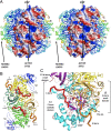
Ribulose 1,5-bisphosphate carboxylase/oxygenase (RuBisCO) is a crucial enzyme in carbon fixation and the most abundant protein on earth. It has been studied extensively by biochemical and structural methods; however, the most essential activation step has not yet been described. Here, we describe the mechanistic details of Lys carbamylation that leads to RuBisCO activation by atmospheric CO(2). We report two crystal structures of nitrosylated RuBisCO from the red algae Galdieria sulphuraria with O(2) and CO(2) bound at the active site. G. sulphuraria RuBisCO is inhibited by cysteine nitrosylation that results in trapping of these gaseous ligands. The structure with CO(2) defines an elusive, preactivation complex that contains a metal cation Mg(2+) surrounded by three H(2)O/OH molecules. Both structures suggest the mechanism for discriminating gaseous ligands by their quadrupole electric moments. We describe conformational changes that allow for intermittent binding of the metal ion required for activation. On the basis of these structures we propose the individual steps of the activation mechanism. Knowledge of all these elements is indispensable for engineering RuBisCO into a more efficient enzyme for crop enhancement or as a remedy to global warming.
Conflict of interest statement
The author declares no conflict of interest.
Figures


Similar articles
-
Ribulose-1,5-bisphosphate carboxylase/oxygenase from thermophilic red algae with a strong specificity for CO2 fixation.Biochem Biophys Res Commun. 1997 Apr 17;233(2):568-71. doi: 10.1006/bbrc.1997.6497. Biochem Biophys Res Commun. 1997. PMID: 9144578
-
Redefinition of rubisco carboxylase reaction reveals origin of water for hydration and new roles for active-site residues.J Am Chem Soc. 2008 Nov 12;130(45):15063-80. doi: 10.1021/ja803464a. Epub 2008 Oct 15. J Am Chem Soc. 2008. PMID: 18855361
-
Effect of Mg2+ on the structure and function of ribulose-1,5-bisphosphate carboxylase/oxygenase.Biol Trace Elem Res. 2008 Mar;121(3):249-57. doi: 10.1007/s12011-007-8050-2. Epub 2007 Oct 30. Biol Trace Elem Res. 2008. PMID: 17968513
-
Structure and function of Rubisco.Plant Physiol Biochem. 2008 Mar;46(3):275-91. doi: 10.1016/j.plaphy.2008.01.001. Epub 2008 Jan 12. Plant Physiol Biochem. 2008. PMID: 18294858 Review.
-
The life of ribulose 1,5-bisphosphate carboxylase/oxygenase--posttranslational facts and mysteries.Arch Biochem Biophys. 2003 Jun 15;414(2):150-8. doi: 10.1016/s0003-9861(03)00122-x. Arch Biochem Biophys. 2003. PMID: 12781766 Review.
Cited by
-
Diel magnesium fluctuations in chloroplasts contribute to photosynthesis in rice.Nat Plants. 2020 Jul;6(7):848-859. doi: 10.1038/s41477-020-0686-3. Epub 2020 Jun 15. Nat Plants. 2020. PMID: 32541951
-
RuBisCO activity assays: a simplified biochemical redox approach for in vitro quantification and an RNA sensor approach for in vivo monitoring.Microb Cell Fact. 2024 Mar 14;23(1):83. doi: 10.1186/s12934-024-02357-6. Microb Cell Fact. 2024. PMID: 38486280 Free PMC article.
-
Carbon Dioxide and the Carbamate Post-Translational Modification.Front Mol Biosci. 2022 Mar 1;9:825706. doi: 10.3389/fmolb.2022.825706. eCollection 2022. Front Mol Biosci. 2022. PMID: 35300111 Free PMC article. Review.
-
TMT-Based Proteomic Analysis of Continuous Cropping Response in Codonopsis tangshen Oliv.Life (Basel). 2023 Mar 13;13(3):765. doi: 10.3390/life13030765. Life (Basel). 2023. PMID: 36983920 Free PMC article.
-
An Insight of RuBisCO Evolution through a Multilevel Approach.Biomolecules. 2021 Nov 25;11(12):1761. doi: 10.3390/biom11121761. Biomolecules. 2021. PMID: 34944405 Free PMC article.
References
-
- Ellis RJ. Most abundant protein in the world. Trends Biochem Sci. 1979;4(11):241–244.
-
- Andersson I. Catalysis and regulation in Rubisco. J Exp Bot. 2008;59(7):1555–1568. - PubMed
-
- Andersson I, Backlund A. Structure and function of Rubisco. Plant Physiol Biochem. 2008;46(3):275–291. - PubMed
-
- Ashida H, et al. RuBisCO-like proteins as the enolase enzyme in the methionine salvage pathway: Functional and evolutionary relationships between RuBisCO-like proteins and photosynthetic RuBisCO. J Exp Bot. 2008;59(7):1543–1554. - PubMed
-
- Lorimer GH, Badger MR, Andrews TJ. The activation of ribulose-1,5-bisphosphate carboxylase by carbon dioxide and magnesium ions. Equilibria, kinetics, a suggested mechanism, and physiological implications. Biochemistry. 1976;15(3):529–536. - PubMed
MeSH terms
Substances
Associated data
- Actions
- Actions
- Actions
LinkOut - more resources
Full Text Sources
Molecular Biology Databases

