. 2012 Nov 13;109(46):E3177-85.
doi: 10.1073/pnas.1213797109. Epub 2012 Oct 29.

Neutrophils orchestrate their own recruitment in murine arthritis through C5aR and FcγR signaling

Affiliations

Neutrophils orchestrate their own recruitment in murine arthritis through C5aR and FcγR signaling

Christian D Sadik et al. Proc Natl Acad Sci U S A. .

Abstract

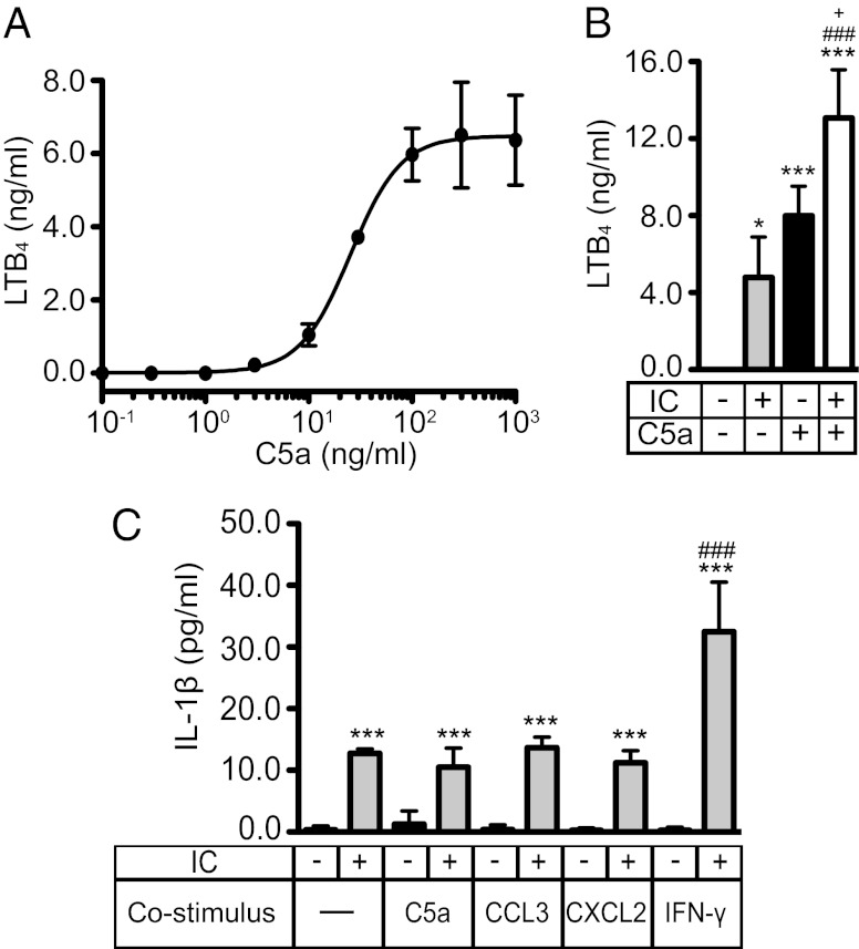
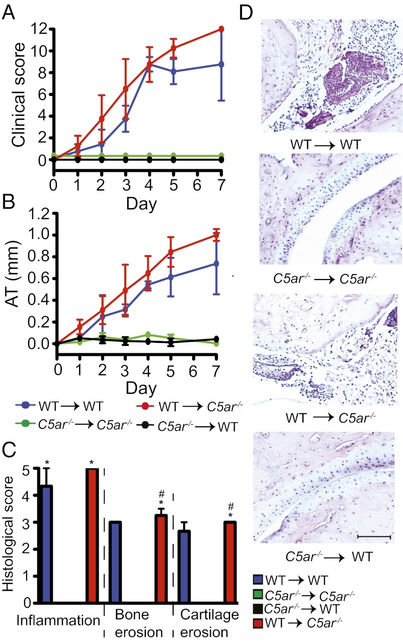
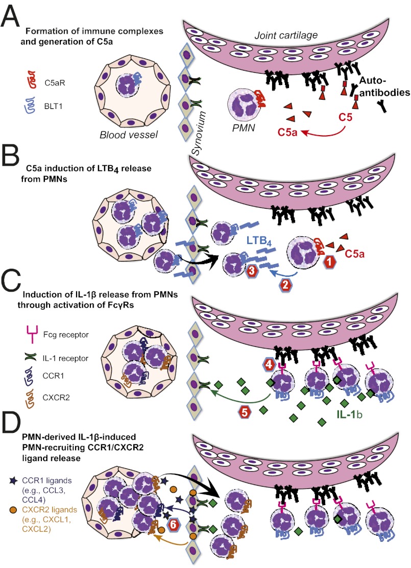
Neutrophil recruitment into the joint is a hallmark of inflammatory arthritides, including rheumatoid arthritis (RA). In a mouse model of autoantibody-induced inflammatory arthritis, neutrophils infiltrate the joint via multiple chemoattractant receptors, including the leukotriene B(4) (LTB(4)) receptor BLT1 and the chemokine receptors CCR1 and CXCR2. Once in the joint, neutrophils perpetuate their own recruitment by releasing LTB(4) and IL-1β, presumably after activation by immune complexes deposited on joint structures. Two pathways by which immune complexes may activate neutrophils include complement fixation, resulting in the generation of C5a, and direct engagement of Fcγ receptors (FcγRs). Previous investigations showed that this model of autoantibody-induced arthritis requires the C5a receptor C5aR and FcγRs, but the simultaneous necessity for both pathways was not understood. Here we show that C5aR and FcγRs work in sequence to initiate and sustain neutrophil recruitment in vivo. Specifically, C5aR activation of neutrophils is required for LTB(4) release and early neutrophil recruitment into the joint, whereas FcγR engagement upon neutrophils induces IL-1β release and subsequent neutrophil-active chemokine production, ensuring continued inflammation. These findings support the concept that immune complex-mediated leukocyte activation is not composed of overlapping and redundant pathways, but that each element serves a distinct and critical function in vivo, culminating in tissue inflammation.


Conflict of interest statement

The authors declare no conflict of interest.

Figures

Fig. 1.
Fig. 1.
C5a and ICs induce LTB4 and IL-1β release from resting PMNs. (A) Dose–response analysis for the release of LTB4 from PMNs after stimulation with varying concentrations of murine recombinant C5a for 1 h. The EC50 of LTB4 induction by C5a is 25.4 ng/mL. (B) Release of LTB4 from PMNs after stimulation with IC, C5a (10 ng/mL), or both combined for 1 h. (C) Release of IL-1β from PMNs after stimulation with IC, C5a (100 ng/mL), CCL3 (100 ng/mL), CXCL2 (100 ng/mL), and IFN-γ (10 ng/mL) alone or combined for 18 h. All data represent concentrations in the supernatant determined by ELISA, shown as mean ± SD (n = 3 independent experiments). *P < 0.05, **P < 0.01, ***P < 0.001 compared with unstimulated control, ### P < 0.001 compared with stimulation with IC, and +P < 0.05 compared with stimulation with C5a.
Fig. 2.
Fig. 2.
C5aR on radiosensitive cells is necessary and sufficient for arthritis. (A and B) Arthritis in BMC of WT and C5ar−/− mice. Clinical score and ankle thickening (AT) were assessed daily (n = 4 mice per group). WT → WT or WT → C5ar−/− vs. C5ar−/−C5ar −/− or C5ar−/− → WT, P < 0.001 in clinical score and in AT. (C) Histological scoring of ankles from mice in A and B. *P < 0.05 compared with C5ar−/−C5ar−/−, and #P < 0.05 compared with C5ar−/− → WT. (D) Respective representative sections. One representative of two independent experiments is shown. Data are presented as mean ± SEM. (Scale bars, 100 μm.)
Fig. 3.
Fig. 3.
5-LO/C5aR but not 5-LO/FcRγ coexpression in radiosensitive cells is required for arthritis. (A and B) Arthritis in WT → WT, Fcer1g−/− → WT, Alox5−/− → WT, Alox5−/−/Fcer1g−/− → WT, Alox5−/−/WT → WT, and Fcer1g−/−/WT → WT BMC (n = 3–4 mice per group; one representative of two independent experiments shown). P < 0.001 in clinical score and AT for WT → WT, Alox5−/−/Fcer1g−/− → WT, Alox5−/−/WT → WT, or Fcer1g−/−/WT → WT vs. Fcer1g−/− → WT, or Alox5−/− → WT. (C) Histological score of ankles from mice in A and B (n = 9–15 mice per group; data compiled from three independent experiments). *P < 0.05, **P < 0.01, and ***P < 0.001 for indicated group vs. Alox5−/− → WT; #P < 0.05, ##P < 0.01, and ###P < 0.001 for indicated group vs. Fcer1g−/− → WT. (D) Respective representative sections. (E and F) Arthritis in WT → WT, C5ar−/− → WT, Alox5−/− → WT, Alox5−/−/C5ar−/− → WT, Alox5−/−/WT → WT, and C5ar−/−/WT → WT BMC (n = 4–5 mice per group; one representative of three independent experiments is shown). P < 0.001 in clinical score for WT → WT, Alox5−/−/WT → WT, or C5ar−/−/WT → WT vs. Alox5−/− → WT, Alox5−/−/C5ar −/− → WT, or C5ar−/− → WT; P < 0.001 in AT for WT → WT or Alox5−/−/WT → WT vs. Alox5−/− → WT, Alox5−/−/C5ar−/− → WT, or C5ar−/− → WT; and P < 0.01 in AT for C5ar−/−/WT → WT vs. Alox5−/− → WT, Alox5−/−/C5ar−/− → WT, or C5ar−/− → WT. (G) Histological score of mice from A and B (n = 9–15 mice per group; compiled from three independent experiments). ***P < 0.001 for indicated group vs. Alox5−/−/C5ar −/− → WT; ##P < 0.01, ###P < 0.001 for indicated group vs. Alox5−/− → WT; ++P < 0.01, +++P < 0.001 for indicated group vs. C5ar−/− → WT. (H) Respective representative sections. All data are presented as mean ± SEM. (Scale bars, 100 μm.)
Fig. 4.
Fig. 4.
C5aR/5-LO coexpression in PMNs is required for arthritis. (A and B) A total of 20 × 106 WT or C5ar−/− PMNs were adoptively transferred into Alox5−/− mice (“WT PMNs formula image 
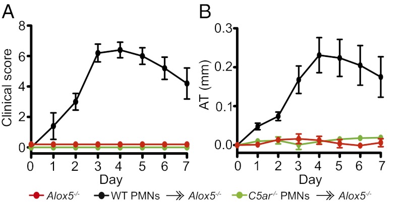
Alox5−/−” and “C5ar−/− PMNs formula image Alox5−/−”) together with K/BxN serum i.v. on days 0 and 2. Alox5−/− mice receiving only K/BxN serum served as controls. Mice were clinically scored daily (n = 4–5 mice per group; one representative of three independent experiments is shown). P < 0.001 for WT PMNs formula image Alox5−/− vs. C5ar−/− PMNs formula image Alox5−/− or Alox5−/− controls in clinical score and ankle thickening. Data are presented as mean ± SEM.
Fig. 5.
Fig. 5.
IL-1β/FcRγ coexpression in PMNs is required for arthritis. (A and B) Arthritis in WT → WT, Fcer1g−/− → WT, Il1a−/−Il1b−/− → WT, Il1a−/−Il1b−/−/Fcer1g−/− → WT, Il1a−/−Il1b−/−/WT → WT, and Fcer1g−/−/WT → WT BMC (n = 4–5 mice per group). P < 0.0001, WT → WT, Il1a−/−Il1b−/−/WT → WT, and Fcer1g−/−/WT → WT vs. Il1a−/−Il1b−/−/Fcer1g−/− → WT, Il1a−/−Il1b−/− → WT, and Fcer1g−/− → WT. (C) Histological score of ankles from mice in A and B (n = 7–9 mice per group; data compiled from two independent experiments). *P < 0.05, **P < 0.01 for indicated group vs. Il1a−/−Il1b−/−/Fcer1g−/− → WT; #P < 0.05, ##P < 0.01 for indicated group vs. Il1a−/−Il1b−/− → WT; +P < 0.05, ++P < 0.01, +++P < 0.001 for indicated group vs. Fcer1g−/− → WT. (D) Respective representative sections. (Scale bars, 100 μm.) (E and F) Clinical evaluation. A total of 20 × 106 WT or Fcer1g−/− PMNs were adoptively transferred i.v. into Il1a−/−Il1b−/− → WT chimeras (WT PMNs formula image 
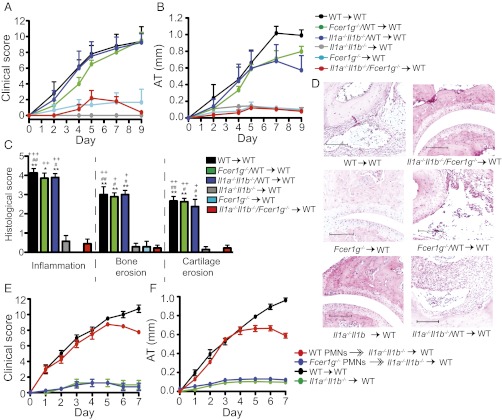
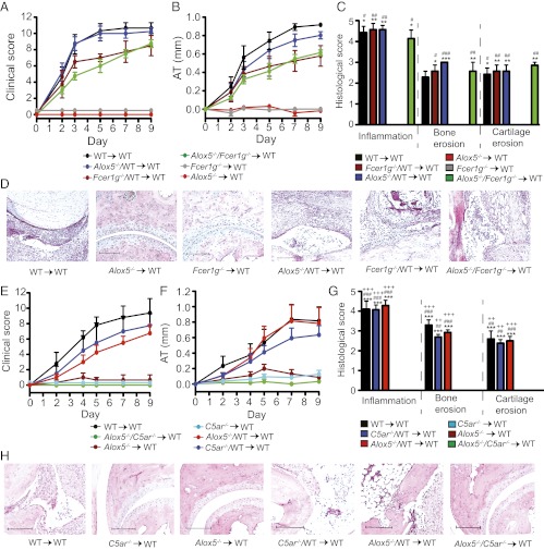
Il1a−/−Il1b−/− → WT; Fcer1g−/− PMNs formula image Il1a−/−Il1b−/− → WT) with K/BxN serum on days 0 and 2. WT → WT and Il1a−/−Il1b−/− → WT chimera obtaining only K/BxN serum served as controls (n = 4 mice per group; one representative of three independent experiments is shown). P < 0.001 for WT PMNs formula image Il1a−/−Il1b−/− → WT vs. Fcer1g−/− PMNs formula image Il1a−/−Il1b−/− → WT or vs. Il1a−/−Il1b−/− → WT chimera in clinical score and AT. All data presented are mean ± SEM.
Fig. 6.
Fig. 6.
FcγRIII is required for IL-1β release. (A and B) Arthritis in WT → WT, Fcgr3−/− → WT, Il1a−/−Il1b−/− → WT, Il1a−/−Il1b−/−/Fcgr3−/− → WT, Il1a−/−Il1b−/−/WT → WT, and Fcgr3−/−/WT → WT BMC (n = 3–5 mice per group). WT → WT, Il1a−/−Il1b−/−/WT → WT, or Fcgr3−/−/WT vs. Il1a−/−Il1b−/−/Fcgr3−/− → WT, Fcgr3−/− → WT, or Il1a−/−Il1b−/− → WT, P < 0.001 in clinical score and AT. One representative of two independent experiments is shown. (C and D) Arthritis in WT → WT, Fcgr3−/− → WT, and Il1a−/−Il1b−/− → WT chimera after i.p. injection of K/BxN serum. Fcgr3−/− → WT and Il1a−/−Il1b−/− → WT chimera were additionally injected i.p. with either 2.5 μg mrIL-1β or its carrier protein (CP) alone, as a control, daily on days 0–2 (n = 3 mice per group). Clinical score and ankle thickening were determined every other day. ***P < 0.001 for Fcgr3−/− → WT + mrIL-1 vs. Fcgr3−/− → WT + CP and Il1a−/−Il1b−/− → WT + mrIL-1 vs. Il1a−/−Il1b−/− → WT + CP in clinical score and AT. All data presented are mean ± SEM.
Fig. P1.
Fig. P1.
Choreography of polymorphonuclear neutrophil (PMN) recruitment in autoantibody-induced arthritis. C5aR and FcγR work in sequence to enable neutrophil-directed neutrophil recruitment. (A) C5a is generated in the joint due to immune complexes deposited on the cartilage surface. (B) C5a activates the first neutrophils in the joint to release LTB4, which in turn recruits a wave of neutrophils into the joint. (C) These neutrophils are activated by immune complexes on the cartilage surface via FcγRIII signaling to release IL-1β, which is a potent activator of chemokine release from synovial cells and endothelial cells in the joint. (D) The release of neutrophil-active chemokines amplifies the recruitment of neutrophils into the joint and enables the perpetuation of arthritis. This nonredundant cascade provides multiple potential therapeutic targets, which are indicated numerically in the schematic, such as 1, C5aR activation; 2, LTB4 generation; 3, BLT1 activation; 4, FcγRIII activation; 5, IL-1R activation; and 6, CCR1 and CXCR2 activation.

References

    1. Sadik CD, Kim ND, Luster AD. Neutrophils cascading their way to inflammation. Trends Immunol. 2011;32(10):452–460. - PMC - PubMed
    1. Sadik CD, Luster AD. Lipid-cytokine-chemokine cascades orchestrate leukocyte recruitment in inflammation. J Leukoc Biol. 2012;91(2):207–215. - PMC - PubMed
    1. Chou RC, et al. Lipid-cytokine-chemokine cascade drives neutrophil recruitment in a murine model of inflammatory arthritis. Immunity. 2010;33(2):266–278. - PMC - PubMed
    1. Kim ND, Chou RC, Seung E, Tager AM, Luster AD. A unique requirement for the leukotriene B4 receptor BLT1 for neutrophil recruitment in inflammatory arthritis. J Exp Med. 2006;203(4):829–835. - PMC - PubMed
    1. Chen M, et al. Neutrophil-derived leukotriene B4 is required for inflammatory arthritis. J Exp Med. 2006;203(4):837–842. - PMC - PubMed

Publication types

MeSH terms