Review
. 2012 Oct 26;338(6106):491-5.
doi: 10.1126/science.1218071.

Glial progenitor cell-based treatment and modeling of neurological disease

Affiliations
Review

Glial progenitor cell-based treatment and modeling of neurological disease

Steven A Goldman et al. Science. .

Abstract

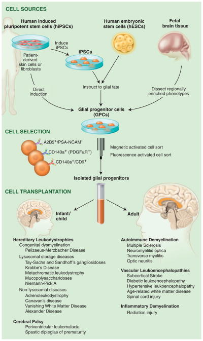
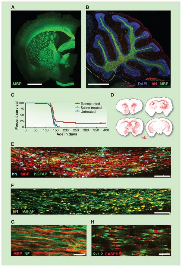
The diseases of myelin are among the most prevalent and disabling conditions in neurology. These diseases include both the vascular and inflammatory demyelinating disorders of adulthood, as well as the childhood leukodystrophies and cerebral palsy. These fundamentally glial disorders may be amenable to treatment by glial progenitor cells (GPCs), which give rise to astroglia and myelin-producing oligodendrocytes. Given the development of new methods for generating and isolating human GPCs, the myelin disorders may now be compelling targets for cell-based therapy. In addition, the efficient engraftment and expansion of human GPCs in murine hosts has led to the development of human glial chimeric mouse brains, which provides new opportunities for studying the species-specific roles of human glia in cognition, as well as in disease pathogenesis.


Figures

Fig. 1
Fig. 1
Glial progenitor cell sources, phenotypes and clinical targets. GPCs may be directly sorted from tissue or generated from either hESCs or hiPSCs and then immunoselected based on their expression of either the A2B5 epitope or CD140a/PDGFαR. The CD140a phenotype includes all potential oligodendrocytes, whereas the tetraspanin CD9 identifies a pro-oligodendrocytic fraction (10). The choice of tissue-, hESC-, or iPSC-derived GPCs depends on whether allogeneic or autologous grafts are desired. Whereas autologous grafts of iPSC-derived GPCs might obviate the need for immunosuppression, their generation may take months, and their use in the hereditary leukodystrophies would first require correction of the underlying genetic disorder in the donor cell pool. At present, such genetic disorders of myelin may be better approached with allografted tissue- or hESC-derived GPCs.
Fig. 2
Fig. 2
Glial progenitor cell graft-mediated myelination of a dysmyelinated host. (A) A 13-month-old shiverer (shi/shi) × rag2−/− mouse, neonatally xenografted with A2B5+/PSA-NCAM hGPCs. MBP, myelin basic protein (shown in green). Shiverer mice do not express MBP; all immunolabeled myelin is of donor origin. (B) Sagittal view through the cerebellum. All cells were stained with 4′,6-diamidino-2-phenylindole (blue); donor cells were identified by human nuclear antigen (hN, red) and MBP (green). (C) Kaplan-Meier plot comparing survival of neonatally engrafted shiverer × rag2−/− mice to untreated and saline-injected controls. (D) CD140a-selected GPCs transplanted into neonatal shiverer × rag2−/− mice expanded and migrated extensively, rendering the brains chimeric. Red dots indicate individual human cells (hN); sacrificed at 3 months. (E and F) Coronal sections of a neonatally engrafted shiverer brain at 3 months, revealing donor-derived myelination (MBP, red) and astrocytic infiltration [human glial fibrillary acidic protein (GFAP), green] of the corpus callosum. (G) Myelin (MBP, red) produced by CD140a-selected hGPCs ensheathes mouse neurofilament-positive axons (NF, green), at 3 months. (H) Reconstituted nodes of Ranvier in the cervical spinal cord of a transplanted and rescued 1-year-old shiverer × rag2−/− mouse, showing paranodal Caspr protein and juxtaparanodal voltage-gated potassium channel protein Kv1.2, symmetrically flanking each axonal node. Untransplanted shiverer brains do not have organized nodes of Ranvier and, hence, cannot support saltatory conduction by central axons (Caspr, red; Kv1.2, green). Scale bars: (A) and (B), 1 mm; (E), 50 μm; (F) and (G), 10 μm; (H), 5 μm. (A) and (B), from (54); (C) and (H), from (5); (D) to (G), from (10).

Similar articles

Cited by

References

    1. Roy N, Windrem M, Goldman SA. In: Myelin Biology and Disorders. Lazzarini R, editor. Elsevier; Amsterdam: 2004. pp. 259–287.
    1. Lee Y, et al. Nature. 2012;487:443. - PMC - PubMed
    1. Tkachev D, et al. Lancet. 2003;362:798. - PubMed
    1. Duncan ID, Brower A, Kondo Y, Curlee JF, Jr, Schultz RD. Proc Natl Acad Sci USA. 2009;106:6832. - PMC - PubMed
    1. Windrem MS, et al. Cell Stem Cell. 2008;2:553. - PMC - PubMed

Publication types