

Gastrointestinal stromal tumor presenting with prominent calcification
- PMID: 23112561
- PMCID: PMC3482655
- DOI: 10.3748/wjg.v18.i39.5645
Gastrointestinal stromal tumor presenting with prominent calcification
Abstract
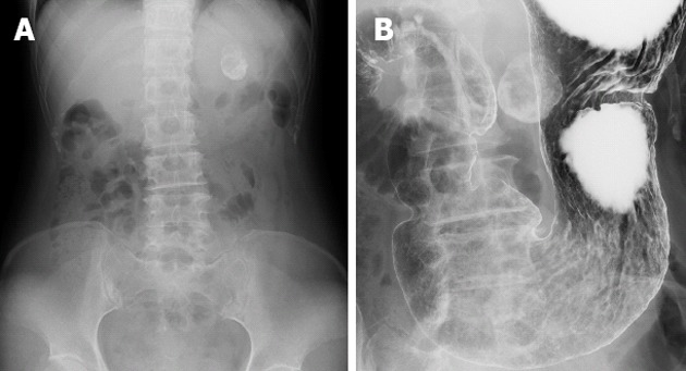
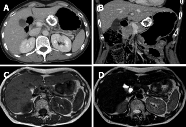
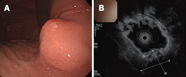
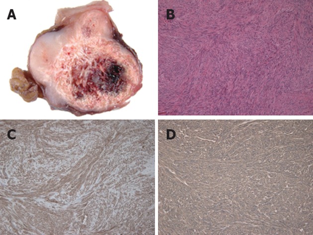
We present a rare case of a gastrointestinal stromal tumor (GIST) in the stomach with prominent calcification at presentation. A 61-year-old woman visited our hospital because of epigastric discomfort. A spherical calcified lesion with a diameter of about 30 mm was incidentally shown in the left upper quadrant on an abdominal X-ray. Computed tomography demonstrated that the tumor was growing from the upper gastric body, with calcification in the peripheral ring area. A laparoscopic partial gastrectomy was performed, and the resected specimen revealed a well-circumscribed tumor with exophytic growth from the gastric muscularis propria. Microscopic examination revealed spindle-shaped tumor cells with calcification and hemorrhage. Additionally, positive immunoreactivity of the tumor to KIT and CD34 and a low mitotic index resulted in the diagnosis of very low risk GIST. There are a few case reports of heavily calcified GIST, although solitary or punctate calcification of primary GIST has been reported in several case series. Dystrophic calcification of necrotic or degenerative tissue is the supposed cause of primary calcified GISTs. In contrast, appearance of calcification after administration of imatinib mesylate, which may be one indicator of disease response, is possibly caused by a different mechanism.
Keywords: Calcification; Computed tomography; Gastrointestinal stromal tumor; Imatinib mesylate; Stomach.
Figures
References
-
- Liegl-Atzwanger B, Fletcher JA, Fletcher CD. Gastrointestinal stromal tumors. Virchows Arch. 2010;456:111–127. - PubMed
-
- Machairas A, Karamitopoulou E, Tsapralis D, Karatzas T, Machairas N, Misiakos EP. Gastrointestinal stromal tumors (GISTs): an updated experience. Dig Dis Sci. 2010;55:3315–3327. - PubMed
-
- Darnell A, Dalmau E, Pericay C, Musulén E, Martín J, Puig J, Malet A, Saigí E, Rey M. Gastrointestinal stromal tumors. Abdom Imaging. 2006;31:387–399. - PubMed
-
- Ghanem N, Altehoefer C, Furtwängler A, Winterer J, Schäfer O, Springer O, Kotter E, Langer M. Computed tomography in gastrointestinal stromal tumors. Eur Radiol. 2003;13:1669–1678. - PubMed
-
- Tateishi U, Hasegawa T, Satake M, Moriyama N. Gastrointestinal stromal tumor. Correlation of computed tomography findings with tumor grade and mortality. J Comput Assist Tomogr. 2003;27:792–798. - PubMed
Publication types
MeSH terms
LinkOut - more resources
Full Text Sources
