Amelioration of endotoxin-induced uveitis treated with an IκB kinase β inhibitor in rats
- PMID: 23112571
- PMCID: PMC3482174
Amelioration of endotoxin-induced uveitis treated with an IκB kinase β inhibitor in rats
Abstract
Purpose: Endotoxin-induced uveitis (EIU) is an animal model for acute ocular inflammation. Several substances play major roles in the development of inflammatory changes in EIU, including tumor necrosis factor-α (TNF-α), interleukin (IL)-1β, and IL-6. These inflammatory cytokines trigger the degradation of IκB by activating IκB kinases (IKKs). Released nuclear factor kappaB (NFκB) subsequently translocates to the nucleus, where NFκB expresses its proinflammatory function. IMD-0354, N-(3,5-Bis-trifluoromethylphenyl)-5-chloro-2-hydroxybenzamide, selectively inhibits IKKβ, particularly when induced by proinflammatory cytokines, such as TNF-α and IL-1β. In the present study, we examined whether IKKβ inhibition has therapeutic effects on EIU by using IMD-0354 and its prodrug IMD-1041.
Methods: Six-week-old male Lewis rats were used. EIU was induced with subcutaneous injections of 200 μg of lipopolysaccharide (LPS) from Escherichia coli that had been diluted in 0.1 ml of phosphate-buffered saline. IMD-0354 was administered intraperitoneally at 30, 10, 3, or 0 mg/kg, suspended in 1.0 ml of 0.5% carboxymethyl cellulose sodium. The prodrug IMD-1041 (100 mg/kg) was also administered orally. The rats were euthanized 24 h after LPS injection, and EIU severity was evaluated histologically. The number of infiltrating cells and the protein, TNF-α, and monocyte chemoattractant protein-1 (MCP-1) concentrations in the aqueous humor were determined. TNF-α and MCP-1 concentrations were quantified with enzyme-linked immunosorbent assay. Eye sections were also stained with anti-NFκB and phosphorylated I-κBα antibodies.
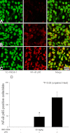
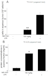
Results: The number of infiltrating cells in aqueous humor was 53.6±9.8×10(5), 72.5±17.0×10(5), 127.25±32.0×10(5), and 132.0±25.0×10(5) cells/ml in rats treated with 30, 10, 3, or 0 mg/kg of IMD-0354, respectively. The total protein concentrations of aqueous humor were 92.6±3.1 mg/ml, 101.5±6.8 mg/ml, 112.6±1.9 mg/ml, and 117.33±1.8 mg/ml in rats treated with 30, 10, 3, and 0 mg/kg of IMD-0354, respectively. Infiltrating cells and protein concentrations were significantly decreased by treatment with IMD-0354 (p<0.01). IMD-0354 treatment significantly reduced the concentration of TNF-α (p<0.05) and MCP-1 (p<0.01) in aqueous humor. The number of NFκB positive nuclei was reduced when treated with IMD-0354. Furthermore, IMD-0354-treated EIU rats showed only background levels of phosphorylated I-κBα; however, it was strongly expressed in the iris-ciliary body cell cytoplasm of the IMD-0354 untreated EIU rats. Oral administration of IMD-1041 also decreased the cell number (p<0.01) and protein concentration (p<0.05) of aqueous humor in EIU.
Conclusions: Acute uveitis was ameliorated by inhibition of IKKβ in rats. IMD-0354 and its prodrug IMD-1041 seem to be promising candidates for treating intraocular inflammation/uveitis.
Figures


References
-
- Rosenbaum JT, McDevitt HO, Guss RB, Egbert PR. Endotoxin-induced uveitis in rats as a model for human disease. Nature. 1980;286:611–3. - PubMed
-
- de Vos AF, van Haren MA, Verhagen C, Hoekzema R, Kijlstra A. Kinetics of intraocular tumor necrosis factor and interleukin-6 in endotoxin-induced uveitis in the rat. Invest Ophthalmol Vis Sci. 1994;35:1100–6. - PubMed
-
- Planck SR, Huang XN, Robertson JE, Rosenbaum JT. Cytokine mRNA levels in rat ocular tissues after systemic endotoxin treatment. Invest Ophthalmol Vis Sci. 1994;35:924–30. - PubMed
-
- Jacquemin E, de Kozak Y, Thillaye B, Courtois Y, Goureau O. Expression of inducible nitric oxide synthase in the eye from endotoxin-induced uveitis rats. Invest Ophthalmol Vis Sci. 1996;37:1187–96. - PubMed
-
- Bellot JL, Palmero M, Garcia-Cabanes C, Espi R, Hariton C, Orts A. Additive effect of nitric oxide and prostaglandin-E2 synthesis inhibitors in endotoxin-induced uveitis in the rabbit. Inflamm Res. 1996;45:203–8. - PubMed
Publication types
MeSH terms
Substances
LinkOut - more resources
Full Text Sources
Research Materials
Miscellaneous
