

The temporal lobes differentiate between the voices of famous and unknown people: an event-related fMRI study on speaker recognition
- PMID: 23112826
- PMCID: PMC3480405
- DOI: 10.1371/journal.pone.0047626
The temporal lobes differentiate between the voices of famous and unknown people: an event-related fMRI study on speaker recognition
Abstract
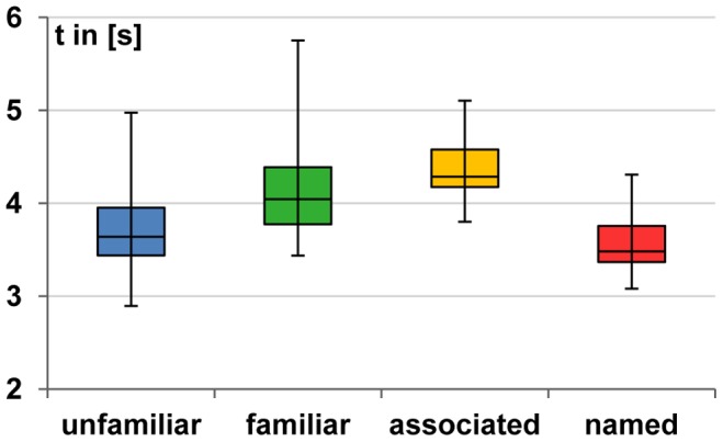
It is widely accepted that the perception of human voices is supported by neural structures located along the superior temporal sulci. However, there is an ongoing discussion to what extent the activations found in fMRI studies are evoked by the vocal features themselves or are the result of phonetic processing. To show that the temporal lobes are indeed engaged in voice processing, short utterances spoken by famous and unknown people were presented to healthy young participants whose task it was to identify the familiar speakers. In two event-related fMRI experiments, the temporal lobes were found to differentiate between familiar and unfamiliar voices such that named voices elicited higher BOLD signal intensities than unfamiliar voices. Yet, the temporal cortices did not only discriminate between familiar and unfamiliar voices. Experiment 2, which required overtly spoken responses and allowed to distinguish between four familiarity grades, revealed that there was a fine-grained differentiation between all of these familiarity levels with higher familiarity being associated with larger BOLD signal amplitudes. Finally, we observed a gradual response change such that the BOLD signal differences between unfamiliar and highly familiar voices increased with the distance of an area from the transverse temporal gyri, especially towards the anterior temporal cortex and the middle temporal gyri. Therefore, the results suggest that (the anterior and non-superior portions of) the temporal lobes participate in voice-specific processing independent from phonetic components also involved in spoken speech material.
Conflict of interest statement
Figures
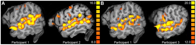
 (Experiment 1, A) or
  (Experiment 2, B) in different participants shown in sagittal plane at
  .
.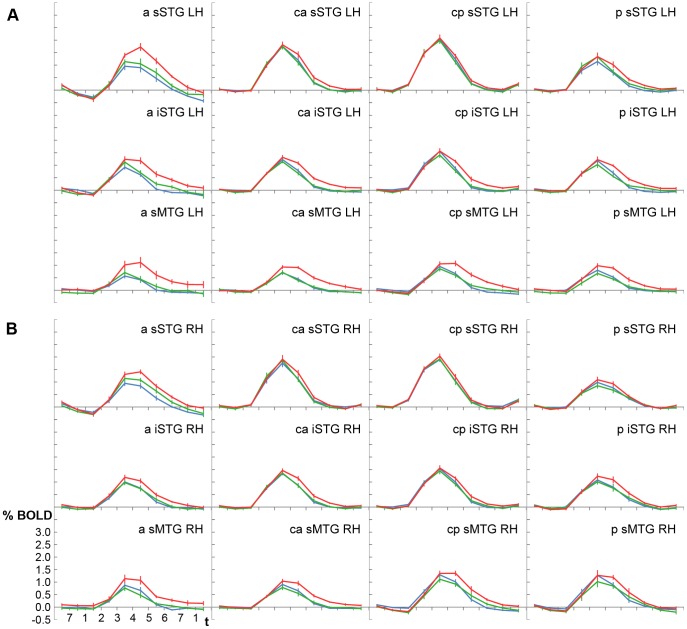
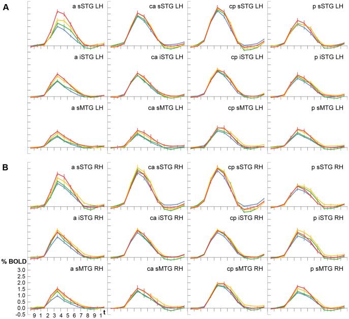
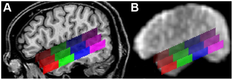
 (right hemisphere). The upper row of ROIs covers the superior part of the STG, the middle row the inferior part of the STG, and the lower row the superior part of the MTG. Red ROIs are located in the anterior temporal lobe, green ROIs in the mid-anterior part, blue ROIs in the mid-posterior part, and purple ROIs in the posterior temporal lobe. The mean Talairach coordinates are given in Table 5. (B) Sagittal view (
 ) of the first functional EPI volume showing those brain regions that produced an MR signal.
References
-
- Belin P, Zatorre RJ (2003) Adaptation to speaker’s voice in right anterior temporal lobe. Neuroreport 14: 2105–2109. - PubMed
-
- Rämä P, Courtney SM (2005) Functional topography of working memory for face or voice identity. NeuroImage 24: 224–234. - PubMed
-
- Shah NJ, Marshall JC, Zafiris O, Schwab A, Zilles K, et al. (2001) The neural correlates of person familiarity: a functional magnetic resonance imaging study with clinical implications. Brain 124: 804–815. - PubMed
-
- Belin P, Zatorre RJ, Ahad P (2002) Human temporal-lobe response to vocal sounds. Cognitive Brain Research 13: 17–26. - PubMed
-
- Meyer M, Zysset S, von Cramon DY, Alter K (2005) Distinct fMRI responses to laughter, speech, and sounds along the human peri-sylvian cortex. Cognitive Brain Research 24: 291–306. - PubMed
Publication types
MeSH terms
LinkOut - more resources
Full Text Sources
Medical
