. 2012;7(10):e47648.
doi: 10.1371/journal.pone.0047648. Epub 2012 Oct 24.

Upregulation of the Tim-3/galectin-9 pathway of T cell exhaustion in chronic hepatitis B virus infection

Affiliations

Upregulation of the Tim-3/galectin-9 pathway of T cell exhaustion in chronic hepatitis B virus infection

Gaia Nebbia et al. PLoS One. 2012.

Abstract

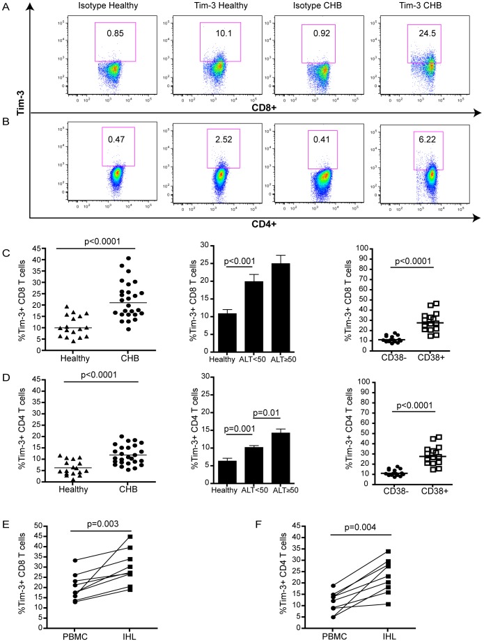
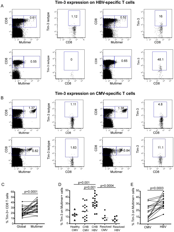
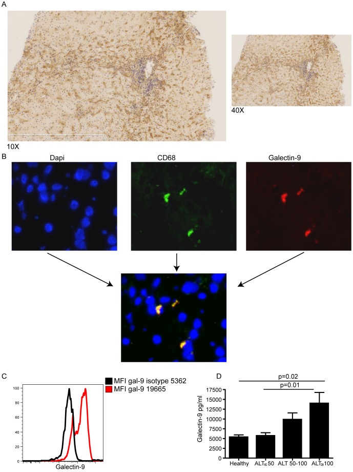
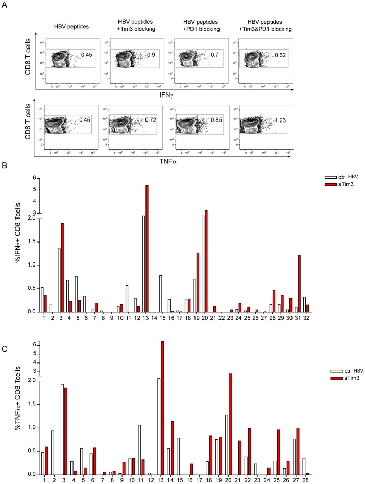
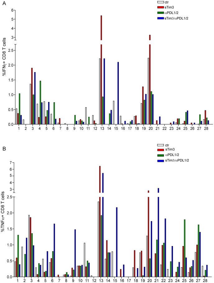
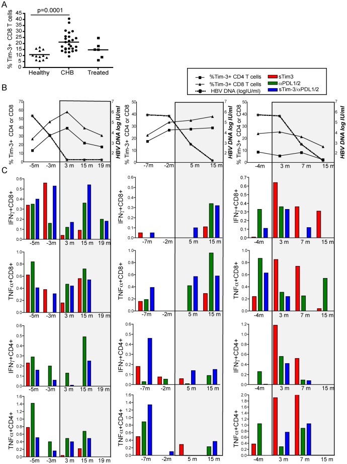
The S-type lectin galectin-9 binds to the negative regulatory molecule Tim-3 on T cells and induces their apoptotic deletion or functional inactivation. We investigated whether galectin-9/Tim-3 interactions contribute to the deletion and exhaustion of the antiviral T cell response in chronic hepatitis B virus infection (CHB). We found Tim-3 to be expressed on a higher percentage of CD4 and CD8 T cells from patients with CHB than healthy controls (p<0.0001) and to be enriched on activated T cells and those infiltrating the HBV-infected liver. Direct ex vivo examination of virus-specific CD8 T cells binding HLA-A2/peptide multimers revealed that Tim-3 was more highly upregulated on HBV-specific CD8 T cells than CMV-specific CD8 T cells or the global CD8 T cell population in patients with CHB (p<0.001) or than on HBV-specific CD8 after resolution of infection. T cells expressing Tim-3 had an impaired ability to produce IFN-γ and TNF-α upon recognition of HBV-peptides and were susceptible to galectin-9-triggered cell death in vitro. Galectin-9 was detectable at increased concentrations in the sera of patients with active CHB-related liver inflammation (p = 0.02) and was strongly expressed by Kupffer cells within the liver sinusoidal network. Tim-3 blockade resulted in enhanced expansion of HBV-specific CD8 T cells able to produce cytokines and mediate cytotoxicity in vitro. Blocking PD-1 in combination with Tim-3 enhanced the number of patients from whom functional antiviral responses could be recovered and/or the strength of responses, indicating that these co-inhibitory molecules play a non-redundant role in driving T cell exhaustion in CHB. Patients taking antivirals able to potently suppress HBV viraemia continued to express Tim-3 on their T cells and respond to Tim-3 blockade. In summary, both Tim-3 and galectin-9 are increased in CHB and may contribute to the inhibition and deletion of T cells as they infiltrate the HBV-infected liver.


Conflict of interest statement

Competing Interests: This work was partially funded by an unrestricted the authors’ adherence to all the PLoS ONE policies on sharing data and materials.

Figures

Figure 1
Figure 1. Tim-3 is up-regulated on peripheral and intrahepatic T cells in HBV infection.
PBMC from 26 HBV infected and 17 uninfected subjects were stained with antibodies against CD3, CD4, CD8, CD38 and Tim-3 or an isotype-matched control Ig. Representative flow cytometry data for (a) CD8 and (b) CD4 T cells from a patient with CHB and a healthy subject. Tim-3 expression on global (c) CD8 and (d) CD4 T cells from healthy individuals and patients with CHB, shown as compiled data and stratified according to ALT level or CD38 co-expression. Expression of Tim-3 on (e) CD8 and (f) CD4 of paired PBMC and IHL from 8 patients with CHB. For all figures, statistical analyses were performed using Mann-Whitney test or the Wilcoxon rank test for paired data; only significant p values (p<0.05) are indicated. Horizontal bars denote medians.
Figure 2
Figure 2. Tim-3 expression is increased on HBV-specific T cells.
HLA-A2+ healthy controls and CHB patients were stained ex vivo with HLA-A2 multimers presenting the CMV-pp65 epitope NLVPMVATV (HLA-A2/NLVP) or a combined panel of 6 HBV-multimers (HLA-A2/core 18–27, envelope 183–191, envelope 348-, envelope 335-,polymerase 455-, polymerase 502-) and with anti-Tim-3 mAb or its isotype. Representative FACS plots from two patients with CHB showing staining for HBV (a) and CMV (b) multimers and Tim-3 expression on gated multimer–specific CD8 T cells compared to an isotype control mAb. (c) Compiled data showing the frequency of virus-specific (multimer+) and global CD8 T cells expressing Tim-3 directly ex vivo in 24 CHB patients. (d) Compiled data showing the frequency of HBV and CMV-specific CD8 T cells expressing Tim-3 directly ex vivo in healthy controls (n = 6), patients with CHB (n = 24) and patients who had resolved HBV (n = 6). (e) Ex-vivo staining in 17 individuals with CHB in whom paired responses could be analysed with both HBV and CMV multimers.
Figure 3
Figure 3. Tim-3 expressing HBV-specific T cells are dysfunctional.
PBMC derived from HLA A2+ patients with CHB were stained with a panel of HLA-A2/HBV multimers and then were stimulated overnight with a pool of HBV peptides of matched specificity to the multimers, followed by intracellular staining for IFN-γor TNF-α. (a) Representative histograms showing levels of Tim-3 (black line) or isotype binding (grey shading) on CD8 T cells binding HLA-A2/HBV peptide multimers or producing IFN-γ upon encounter with HBV peptides. (b) Compiled data from 10 patients with CHB. (c) Tim-3 expression on CD8 T cells binding HLA-A2/HBV peptide multimers or producing TNFa upon stimulation with HBV peptides. (d) FACS plots and (e) summary data showing the induction of caspases (FLICA) and 7AAD in CD8 and CD4 T cells with or without the addition of galectin-9. Active caspases, indicating apoptosis, were determined using a fluorescent-labelled inhibitor of polycaspases (FAM-VAD-FMK, FLICA), and death was identified by 7AAD stain. Early apoptotic events are indicated in the lower right quadrant (FLICA+7AAD−), late apoptotic events in the right upper quadrant (FLICA+7AAD+) and necrotic cells in the left upper quadrant (7AAD+FLICA-). ‘Total death’ was estimated by summing events in these 3 quadrants.
Figure 4
Figure 4. The Tim-3 ligand galectin-9 is expressed by Kupffer cells, and its secretion is increased in active CHB.
Immunohistochemistry of a section from a cryopreserved CHB liver biopsy stained with a polyclonal galectin-9 antibody (shown in brown) at 10X and 40X magnification. (b) Immunofluorescence of a section from a cryopreserved CHB liver biopsy stained with DAPI (blue, left panel), anti-CD68 mAb (green, central panel) and galectin 9 polyclonal antibody (red, right panel). Double positive staining is indicated in yellow in the lower panel. (c) Kupffer cells were isolated from 4 liver explants and stained with CD14, CD68 and galectin-9 or its isotype. Representative histograms show galectin-9 staining on CD14+CD68+ fraction compared to isotype-matched control. (d) Galectin-9 levels were analysed in the serum of 10 healthy subjects and 42 CHB patients: 16 with ALT<50, 17 with ALT 50–100 and 9 with ALT >100 (mean and SEM shown).
Figure 5
Figure 5. Blocking the Tim-3 pathway can increase the frequency of IFNγ and TNFα-producing HBV-specific CD8 T cells.
(a) Representative dot plot showing recovery of HBV-specific CD8 T cells responses (IFN-γ, TNF-α) in a patient with CHB. PBMC were stimulated with peptides and cultured for 10 days in the presence of soluble Tim-3 FC chimera, PDL1/L2 blocking antibody or both. Summary data of the effect of blocking Tim-3 on IFN-γ (b) and TNF-α (c) production by CD8 T cells in response to HBV peptides.
Figure 6
Figure 6. Non-redundant roles for Tim-3 and PD-L1/2 blockade in recovery of functional HBV-specific T cell responses.
Summary data from 28 patients with CHB of the percent of HBV-specific CD8 T cells producing IFN-γ (a) or TNF-α (b) upon blockade of Tim-3 (red bars), PDL1/L2 (green bars) or dual Tim-3 and PDL1/L2 (blue bars), compared to stimulation with peptides without blocking (white bars).
Figure 7
Figure 7. Effect of antiviral treatment on Tim-3 expression and response to Tim-3 blockade.
(a) Tim-3 expression on global CD3 CD4+ T cells and CD3 CD8+ T cells. Each dot represents an individual data point (12 healthy, 26 CHB, 6 CHB on antivirals with undetectable viral load); horizontal lines represent the mean. (b) Percent of CD4 (square), CD8 (triangle) T cells expressing Tim-3 (left y axis) and HBV load circle, right y axis) plotted longitudinally for 3 patients starting antiviral therapy. (c) PBMC sampled at the indicated time points from these three individuals were stimulated for ten days with HBV OLP with control IgG, Tim-3 Fc chimera, anti-PDL1/PDL2 antibodies or both. Bars represent the % of HBV-specific CD8 T cells producing IFN-γ or TNF-α following blockade after subtracting the frequency detectable without any blockade.

Similar articles

Cited by

References

    1. Rabinovich GA, Toscano MA (2009) Turning ‘sweet’ on immunity: galectin-glycan interactions in immune tolerance and inflammation. Nat Rev Immunol 9: 338–352. - PubMed
    1. Wada J, Ota K, Kumar A, Wallner EI, Kanwar YS (1997) Developmental regulation, expression, and apoptotic potential of galectin-9, a beta-galactoside binding lectin. J Clin Invest 99: 2452–2461. - PMC - PubMed
    1. Monney L, Sabatos CA, Gaglia JL, Ryu A, Waldner H, et al. (2002) Th1-specific cell surface protein Tim-3 regulates macrophage activation and severity of an autoimmune disease. Nature 415: 536–541. - PubMed
    1. Zhu C, Anderson AC, Schubart A, Xiong H, Imitola J, et al. (2005) The Tim-3 ligand galectin-9 negatively regulates T helper type 1 immunity. Nat Immunol 6: 1245–1252. - PubMed
    1. Golden-Mason L, Palmer BE, Kassam N, Townshend-Bulson L, Livingston S, et al. (2009) Negative immune regulator Tim-3 is overexpressed on T cells in hepatitis C virus infection and its blockade rescues dysfunctional CD4+ and CD8+ T cells. J Virol 83: 9122–9130. - PMC - PubMed

Publication types

MeSH terms