. 2012 Oct 31:12:246.
doi: 10.1186/1471-2180-12-246.

Macropinocytosis is responsible for the uptake of pathogenic and non-pathogenic mycobacteria by B lymphocytes (Raji cells)

Affiliations

Macropinocytosis is responsible for the uptake of pathogenic and non-pathogenic mycobacteria by B lymphocytes (Raji cells)

Blanca Estela García-Pérez et al. BMC Microbiol. .

Abstract

Background: The classical roles of B cells include the production of antibodies and cytokines and the generation of immunological memory, these being key factors in the adaptive immune response. However, their role in innate immunity is currently being recognised. Traditionally, B cells have been considered non-phagocytic cells; therefore, the uptake of bacteria by B cells is not extensively documented. In this study, we analysed some of the features of non-specific bacterial uptake by B lymphocytes from the Raji cell line. In our model, B cells were infected with Mycobacterium tuberculosis (MTB), Mycobacterium smegmatis (MSM), and Salmonella typhimurium (ST).

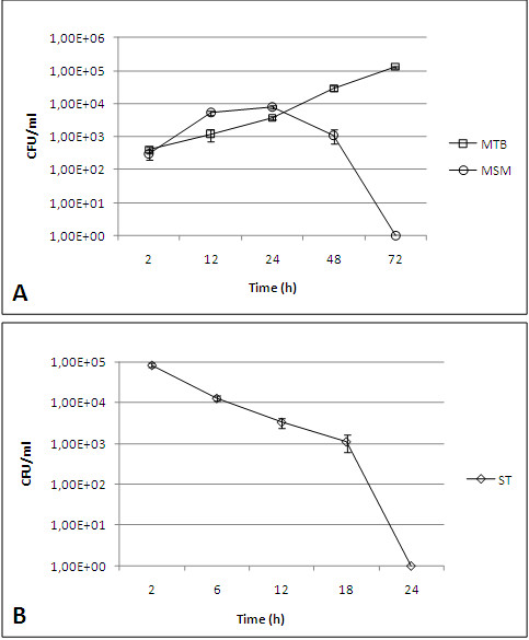
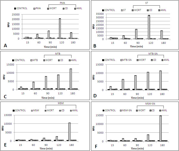
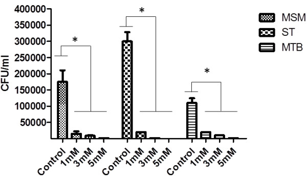
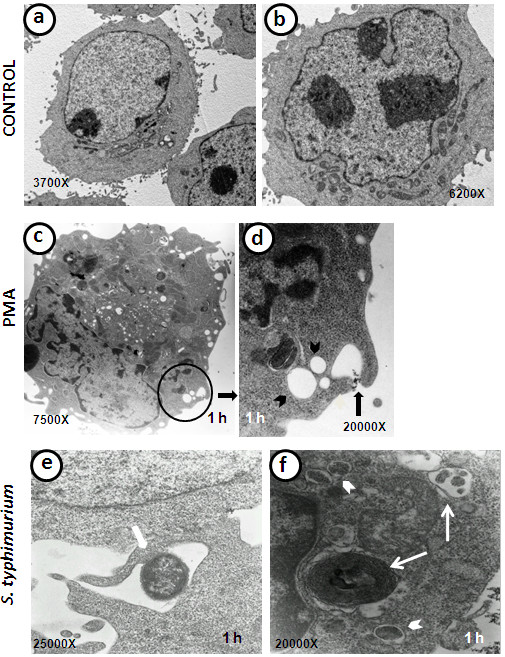
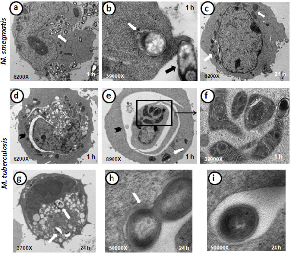
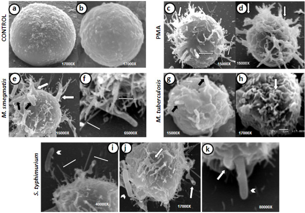
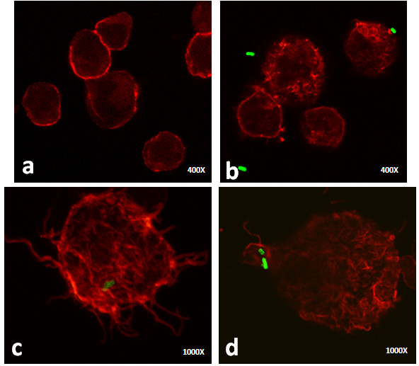
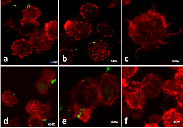
Results: Our observations revealed that the Raji B cells were readily infected by the three bacteria that were studied. All of the infections induced changes in the cellular membrane during bacterial internalisation. M. smegmatis and S. typhimurium were able to induce important membrane changes that were characterised by abundant filopodia and lamellipodia formation. These membrane changes were driven by actin cytoskeletal rearrangements. The intracellular growth of these bacteria was also controlled by B cells. M. tuberculosis infection also induced actin rearrangement-driven membrane changes; however, the B cells were not able to control this infection. The phorbol 12-myristate 13-acetate (PMA) treatment of B cells induced filopodia and lamellipodia formation, the production of spacious vacuoles (macropinosomes), and the fluid-phase uptake that is characteristic of macropinocytosis. S. typhimurium infection induced the highest fluid-phase uptake, although both mycobacteria also induced fluid uptake. A macropinocytosis inhibitor such as amiloride was used and abolished the bacterial uptake and the fluid-phase uptake that is triggered during the bacterial infection.

Conclusions: Raji B cells can internalise S. typhimurium and mycobacteria through an active process, such as macropinocytosis, although the resolution of the infection depends on factors that are inherent in the virulence of each pathogen.


Figures

Figure 1
Figure 1
Colony forming units (CFU) of S. typhimurium and mycobacteria in B cells. a) Time-dependent CFU counts of intracellular M. smegmatis (MSM) (circles) and M. tuberculosis (MTB) (squares). The growth of M. smegmatis is controlled by the end of the kinetics, whereas M. tuberculosis survives and multiplies. b) Time-dependent CFU counts of intracellular S. typhimurium (ST). The intracellular growth was rapidly controlled by the B cells compared to the mycobacteria. Each point represents the mean ± standard error (SE) of triplicate measurements. The experiment presented is representative of three independent repetitions.
Figure 2
Figure 2
Fluid-phase uptake by Raji B cells induced by different treatments. B cells were infected with M. tuberculosis (MTB), M. smegmatis (MSM), and S. typhimurium (ST), or treated with phorbol 12-myristate 3-acetate (PMA), M. tuberculosis culture supernatant (MTB-SN), or M. smegmatis culture supernatant (MSM-SN). The fluorescent fluid-phase uptake was determined by the quantification of the relative fluorescence units (RFU) at several time points (15, 60, 90, 120, and 180 min). B cells that were not treated served as the control (CONTROL) for each treatment. The effect of several inhibitors on the fluid-phase uptake was also monitored. Each of the inhibitors (cytochalasin (CD), wortmannin (WORT), and amiloride (AMIL) was individually added to the following treatments/infections: a) PMA treatment, b) ST, c) MTB, d) MTB-SN, e) MSM, f) MSM-SN. Each bar represents the mean of four different measurements. There were statistically significant differences (p <0.01) when the infected, PMA-treated and SN-treated B cells were compared with i) the control cells, ii) the infected cells in the presence of the inhibitors, and iii) the PMA-treated or SN-treated cells in the presence of the inhibitors. The experiment presented is representative of three independent repetitions.
Figure 3
Figure 3
Bacterial uptake by Raji B cells is inhibited by amiloride treatment. B cells were infected with M. tuberculosis (MTB), M. smegmatis (MSM), and S. typhimurium (ST) for 90 min. The cells were treated with 1, 3 or 5 mM amiloride before and during the infection. The CFU counts were determined and recorded; * statistically significant differences (p <0.01) when the untreated/infected cells were compared with amiloride-treated/infected cells.
Figure 4
Figure 4
Ultrastructure of B cells infected with S. typhimurium (ST) and stimulated with phorbol 12-myristate 3-acetate (PMA). a-b) Control B cells. c) PMA-stimulated B cell, which has abundant vacuoles of different sizes. d) The field magnification of a PMA-stimulated B cell (circle) shows macropinosome formation (black narrow) and the presence of macropinosomes that are already formed in various sizes (arrowheads). e) Micrograph of S. typhimurium-infected B cell, which shows that the bacillus is surrounded by large membrane extensions (narrow). f) S. typhimurium-infected B cell with internalised bacteria (arrowheads), thin narrows depicts a multilamellar structure (left) and a late degradative autophagic vacuole (LDAV) (right).
Figure 5
Figure 5
Ultrastructure of B cells infected with M. smegmatis (MSM) and M. tuberculosis (MTB). a) MSM-infected B cell with abundant internalised bacilli (white arrow) after 1 h of infection. b) MSM-infected B cell after 1 h of infection, which shows the binding of a bacillus to a lamellipodium (black arrow) and the destruction of an intracellular bacillus contained in a vacuole (white arrow). c) MSM-infected B cell at 24 h post-infection, which shows that the cell morphology was recovered and that no internalised bacilli were present, although some swollen mitochondria were still observed (white arrows). d-e) After 1 h of infection, a B cell infected with MTB exhibits a large number of alterations, abundant vacuoles, swollen mitochondria, internalised mycobacteria (white arrow), and “curved vacuoles” (black arrowheads). f) Magnification of a B cell infected with MTB (square), which shows that some of the altered mitochondria are in the process of forming double to multi-membrane vacuoles (autophagy-like vacuoles). g) B cell infected with MTB for 24 h shows intracellular bacilli in vacuoles (white arrows), abundant vacuoles, and an electro-dense cellular nucleus, which suggests strong damage. h) Replicating mycobacteria in spacious vacuole (white arrow) formed in a B cell infected with MTB for 24 h. g) Detail of MTB bacillus in a spacious vacuole after 24 h of B cell infection.
Figure 6
Figure 6
Scanning electron micrographs of B cells infected with mycobacteria or S. typhimurium (ST) or treated with phorbol 12-myristate 3-acetate (PMA). a-b) Non-infected B cells. c-d) PMA-treated B cells, which exhibit abundant long, thin, and wide membrane extensions that resemble filopodia (thin arrows) and lamellipodia (wide arrows). e-f) B cells infected with M. smegmatis (MSM) show abundant membrane filopodia (thin white narrows) and lamellipodia formation (wide white narrows) and attached bacilli that are trapped by the membrane projections (black arrows). g-h) Mycobacterium tuberculosis (MTB)-infected B cells show membrane ruffling (white arrow) and some bacilli bound to the cell (black arrows). i-k) S. typhimurium-infected B cells show filopodia (thin white arrows) and lamellipodia formation (wide white arrows). The white arrowheads depict attached bacilli and a bacillus that is surrounded by forming lamellipodia.
Figure 7
Figure 7
Confocal images of uninfected and S. typhimurium (ST)-infected B cells. The actin filaments were labelled with rhodamine-phalloidin and the bacteria were stained with fluorescein isothiocyanate (FITC). a) Uninfected cells present peripheral and homogeneous fluorescent staining. b) One h after infection, S. typhimurium induced actin cytoskeletal rearrangements that are responsible for membrane ruffling; in addition, a bacillus that is attached to these structures was observed (upper right corner). c-d) After 3 h of infection, longer actin projections and actin redistribution were observed, and some bacilli were found inside the B cell (c) or surrounded by actin organisations (d).
Figure 8
Figure 8
Confocal images of B cells infected with mycobacteria. The actin filaments were labelled with rhodamine-phalloidin and the bacteria were stained with fluorescein isothiocyanate (FITC). a) M. smegmatis (MSM) infection caused evident actin rearrangements within 1 h of infection; a mycobacterium was observed attached to the cell. b-c) After 3 h of infection with MSM, intracellular bacteria were observed (b) and long actin filaments were evident (c). d) M. tuberculosis (MTB) infection induced actin reorganisation after 1 h of infection, and bacilli attached to the cells were observed; e-f) B cells, after 3 h of infection with MTB, presented actin cytoskeletal changes in cells without any adhered or intracellular bacteria.

References

    1. Lund FE, Garvy BA, Randall TD, Harris DP. Regulatory roles for cytokine-producing B cells in infection and autoimmune disease. Curr Dir Autoimmun. 2005;8:25–54. - PubMed
    1. Batista FD, Iber D, Neuberger MS. B cells acquire antigen from target cells after synapse formation. Nature. 2001;411:489–494. doi: 10.1038/35078099. - DOI - PubMed
    1. Gupta N, DeFranco AL. Lipid rafts and B cell signaling. Semin Cell Dev Biol. 2007;18:616–626. doi: 10.1016/j.semcdb.2007.07.009. - DOI - PMC - PubMed
    1. Putnam MA, Moquin AE, Merrihew M, Outcalt C, Sorge E, Caballero A, Gondré-Lewis TA, Drake JR. Lipid raft-independent B cell receptor-mediated antigen internalization and intracellular trafficking. J Immunol. 2003;170:905–912. - PubMed
    1. Chiron D, Bekeredjian-Ding I, Pellat-Deceunynck C, Bataille R, Jego G. Toll-like receptors: lessons to learn from normal and malignant human B cells. Blood. 2008;112:2205–2213. doi: 10.1182/blood-2008-02-140673. - DOI - PMC - PubMed

Publication types

LinkOut - more resources