Comparative Study
. 2013 Jan;92(1):51-7.
doi: 10.1177/0022034512466263. Epub 2012 Oct 31.

Endothelial differentiation of SHED requires MEK1/ERK signaling

Affiliations
Comparative Study

Endothelial differentiation of SHED requires MEK1/ERK signaling

L W Bento et al. J Dent Res. 2013 Jan.

Abstract

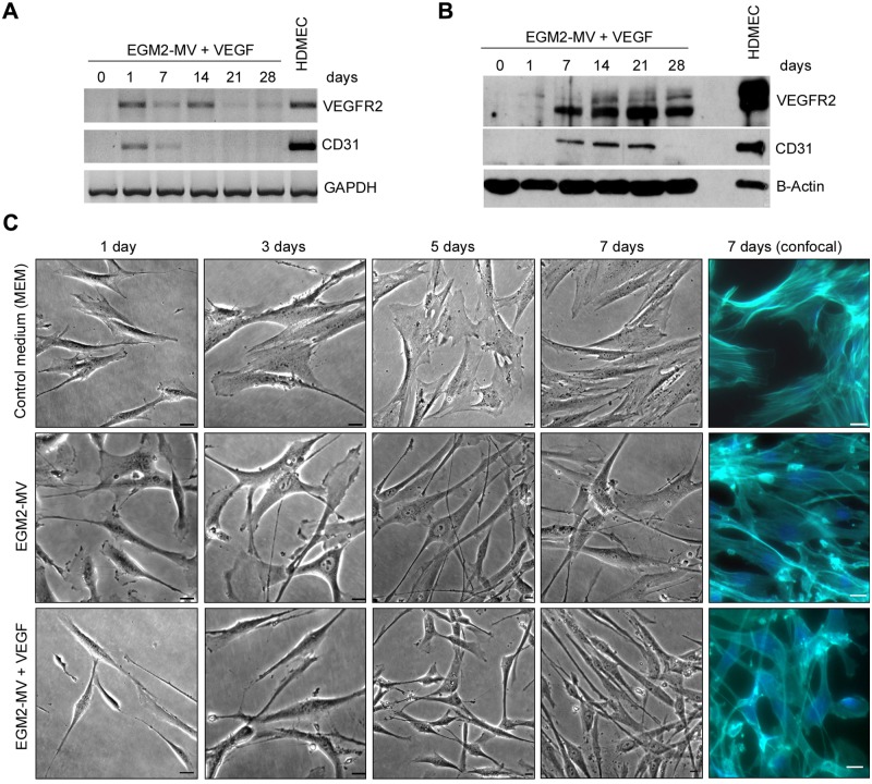
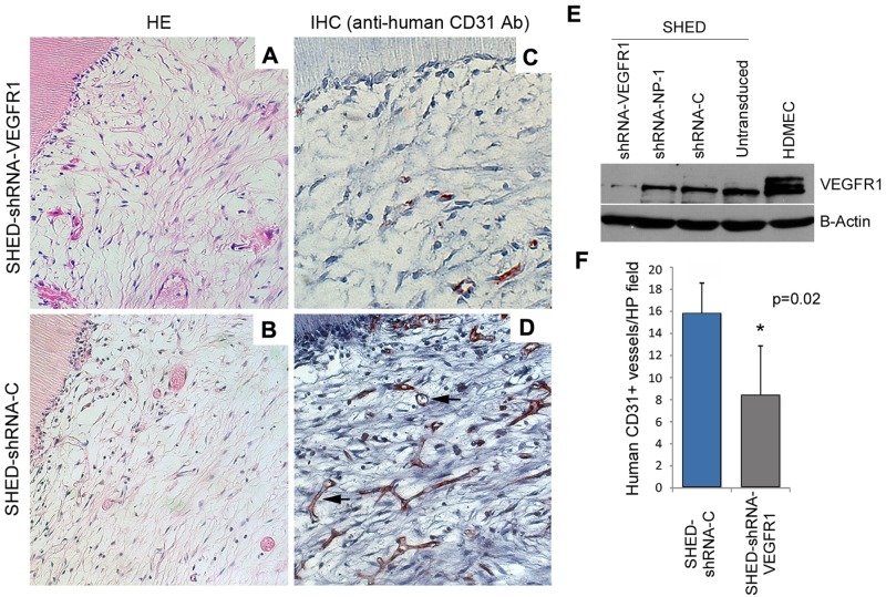
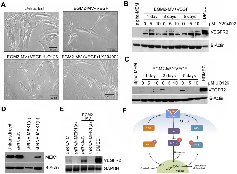
The discovery that dental pulp stem cells are capable of differentiating into endothelial cells raises the exciting possibility that these cells can be a single source of odontoblasts and vascular networks in dental tissue engineering. The purpose of this study was to begin to define signaling pathways that regulate endothelial differentiation of SHED. Stem cells from exfoliated deciduous teeth (SHED) exposed to endothelial growth medium (EGM-2MV) supplemented with vascular endothelial growth factor (VEGF) differentiated into VEGFR2-positive and CD31-positive endothelial cells in vitro. In vivo, VEGFR1-silenced SHED seeded in tooth slice/ scaffolds and transplanted into immunodeficient mice showed a reduction in human CD31-positive blood vessels as compared with controls (p = 0.02). Exposure of SHED to EGM2-MV supplemented with VEGF induced potent activation of ERK and Akt signaling, while it inhibited phosphorylation of STAT3. Notably, genetic (MEK1 silencing) or chemical (U0126) inhibition of ERK signaling restored constitutive STAT3 phosphorylation and inhibited the differentiation of SHED into endothelial cells. Collectively, analysis of these data unveiled the VEGF/MEK1/ERK signaling pathway as a key regulator of the endothelial differentiation of dental pulp stem cells.


Conflict of interest statement

The authors declare no potential conflicts of interest with respect to the authorship and/or publication of this article.

Figures

Figure 1.
Figure 1.
Time-course of the differentiation of SHED into endothelial cells. (A) Time-course of mRNA and (B) protein expression of endothelial cell markers VEGFR2 and CD31 upon culture of SHED cells with EGM2-MV + 50 ng/mL rhVEGF165. (C) Cell morphology over time upon culture of SHED cells with basal medium (alpha-MEM), endothelial growth medium (EGM2-MV), or EGM2-MV supplemented with 50 ng/mL rhVEGF165 (bar = 20 µm).
Figure 2.
Figure 2.
VEGFR1 silencing inhibits endothelial differentiation of SHED in vivo. (A, C) Tooth slice/scaffolds seeded with VEGFR1-silenced SHED (SHED-shRNA-VEGFR1) and (B, D) vector control SHED (SHED-shRNA-C) were transplanted into the subcutaneous space of immunodeficient mice. After 28 days, tooth slice/scaffolds were retrieved, fixed, and analyzed by hematoxylin-eosin staining immunohistochemistry with anti-human CD31. Black arrows indicate CD31-positive blood vessels. (E) Western blot to evaluate the effectiveness of VEGFR1 knockdown in SHED cells. (F) Microvessel density analysis from 4 tooth slice/scaffolds and 5 microscopic fields/specimen (at 200x) per experimental condition.
Figure 3.
Figure 3.
Activation of signaling upon exposure of SHED to differentiation medium. (A) Western blot depicting effect of differentiation medium (EGM-2MV + rhVEGF165) on phosphorylated and total ERK, Akt, and STAT3. (B) Western blot showing the effects of inhibitors for ERK (U0126), Akt (LY294002), or STAT3 (Stattic V) on signaling induced by the differentiation medium.
Figure 4.
Figure 4.
MEK1/ERK signaling is required for endothelial differentiation of SHED in vitro. (A) Qualitative analysis of the morphology of SHED cells treated with differentiation medium in the presence of U0126 or LY294002. (B) Western blot showing the effect of Akt inhibition with LY294002 or (C) the effect of ERK inhibition with U0126 on endothelial cell differentiation of SHED, as determined by VEGFR2 expression. (D) Effects of 2 different shRNA-MEK1 sequences (a,b) on silencing of MEK1 in SHED cells. (E) Effect of MEK1 silencing on endothelial cell differentiation of SHED, as determined by VEGFR2 expression. (F) Schematic diagram depicting the signaling pathways studied here.

References

    1. Albelda SM, Muller WA, Buck CA, Newman PJ.(1991). Molecular and cellular properties of PECAM-1 (endoCAM/CD31): a novel vascular cell-cell adhesion molecule. J Cell Biol 114:1059-1068. - PMC - PubMed
    1. Behr B, Tang C, Germann G, Longaker MT, Quarto N. (2011). Locally applied vascular endothelial growth factor A increases the osteogenic healing capacity of human adipose-derived stem cells by promoting osteogenic and endothelial differentiation. Stem Cells 29:286-296. - PMC - PubMed
    1. Cao Y. (2009). Positive and negative modulation of angiogenesis by VEGFR1 ligands. Sci Signal 2(59):re1. - PubMed
    1. Cao Y, Sun Z, Liao L, Meng Y, Han Q, Zhao RC. (2005). Human adipose tissue-derived stem cells differentiate into endothelial cells in vitro and improve postnatal neovascularization in vivo. Biochem Biophys Res Commun 332:370-379. - PubMed
    1. Casagrande L, Demarco FF, Zhang Z, Araujo FB, Shi S, Nör JE. (2010). Dentin-derived BMP-2 and odontoblast differentiation. J Dent Res 89:603-608. - PubMed

Publication types

MeSH terms