

Implications for the mammalian sialidases in the physiopathology of skeletal muscle
- PMID: 23114189
- PMCID: PMC3534598
- DOI: 10.1186/2044-5040-2-23
Implications for the mammalian sialidases in the physiopathology of skeletal muscle
Abstract
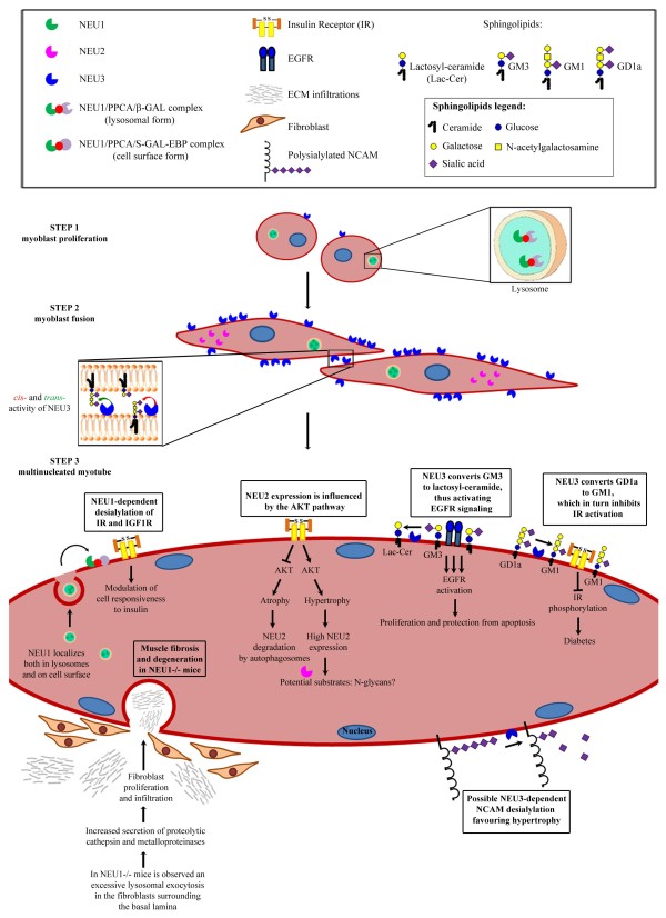
The family of mammalian sialidases is composed of four distinct versatile enzymes that remove negatively charged terminal sialic acid residues from gangliosides and glycoproteins in different subcellular areas and organelles, including lysosomes, cytosol, plasma membrane and mitochondria. In this review we summarize the growing body of data describing the important role of sialidases in skeletal muscle, a complex apparatus involved in numerous key functions and whose functional integrity can be affected by various conditions, such as aging, chronic diseases, cancer and neuromuscular disorders. In addition to supporting the proper catabolism of glycoconjugates, sialidases can affect different signaling pathways by desialylation of many receptors and modulation of ganglioside content in cell membranes, thus actively participating in myoblast proliferation, differentiation and hypertrophy, insulin responsiveness and skeletal muscle architecture.
Figures
Similar articles
-
Downregulation of Zebrafish Cytosolic Sialidase Neu3.2 Affects Skeletal Muscle Development.Int J Mol Sci. 2023 Sep 1;24(17):13578. doi: 10.3390/ijms241713578. Int J Mol Sci. 2023. PMID: 37686385 Free PMC article.
-
Sialidases in vertebrates: a family of enzymes tailored for several cell functions.Adv Carbohydr Chem Biochem. 2010;64:403-79. doi: 10.1016/S0065-2318(10)64007-3. Adv Carbohydr Chem Biochem. 2010. PMID: 20837202 Review.
-
Biological and Pathological Roles of Ganglioside Sialidases.Prog Mol Biol Transl Sci. 2018;156:121-150. doi: 10.1016/bs.pmbts.2017.12.005. Epub 2018 Jan 9. Prog Mol Biol Transl Sci. 2018. PMID: 29747812 Review.
-
Recent development in mammalian sialidase molecular biology.Neurochem Res. 2002 Aug;27(7-8):649-63. doi: 10.1023/a:1020276000901. Neurochem Res. 2002. PMID: 12374200 Review.
-
Mammalian sialidases: physiological and pathological roles in cellular functions.Glycobiology. 2012 Jul;22(7):880-96. doi: 10.1093/glycob/cws057. Epub 2012 Feb 28. Glycobiology. 2012. PMID: 22377912 Review.
Cited by
-
Sialic acids in the brain: gangliosides and polysialic acid in nervous system development, stability, disease, and regeneration.Physiol Rev. 2014 Apr;94(2):461-518. doi: 10.1152/physrev.00033.2013. Physiol Rev. 2014. PMID: 24692354 Free PMC article. Review.
-
Neuraminidase-1 mediates skeletal muscle regeneration.Biochim Biophys Acta. 2015 Sep;1852(9):1755-64. doi: 10.1016/j.bbadis.2015.05.006. Epub 2015 May 19. Biochim Biophys Acta. 2015. PMID: 26001931 Free PMC article.
-
Sialometabolism in Brain Health and Alzheimer's Disease.Front Neurosci. 2021 Mar 30;15:648617. doi: 10.3389/fnins.2021.648617. eCollection 2021. Front Neurosci. 2021. PMID: 33867926 Free PMC article. Review.
-
Inhibitors of the Sialidase NEU3 as Potential Therapeutics for Fibrosis.Int J Mol Sci. 2022 Dec 23;24(1):239. doi: 10.3390/ijms24010239. Int J Mol Sci. 2022. PMID: 36613682 Free PMC article. Review.
-
Serum Sialylation Changes in Actinic Keratosis and Cutaneous Squamous Cell Carcinoma Patients.J Pers Med. 2021 Oct 15;11(10):1027. doi: 10.3390/jpm11101027. J Pers Med. 2021. PMID: 34683168 Free PMC article.
References
-
- Saito M, Yu RK. In: Biology of the Sialic Acids. Rosenberg A, editor. Plenum Press; 1995. Biochemistry and function of sialidases; pp. 261–313.
-
- Monti E, Bonten E, D'Azzo A, Bresciani R, Venerando B, Borsani G, Schauer R, Tettamanti G. Sialidases in vertebrates: a family of enzymes tailored for several cell functions. Adv Carbohydr Chem Biochem. 2010;64:403–479. - PubMed
-
- Schauer R, Kamerling JP. In: Glycoproteins II. Montreuil J, Vliegenthart JF, Schachte H, editor. Amsterdam: Elsevier Science B.V; 1997. Chemistry, biochemistry and biology of sialic acids; pp. 243–402.
-
- Miyagi T, Konno K, Emori Y, Kawasaki H, Suzuki K, Yasui A, Tsuik S. Molecular cloning and expression of cDNA encoding rat skeletal muscle cytosolic sialidase. J Biol Chem. 1993;268:26435–26440. - PubMed
LinkOut - more resources
Full Text Sources
